Stem borer (Diptera: Agromyzidae) in Clematis [0159]

OrderDiptera
FamilyAgromyzidae[M,L,P,A]
Lower taxonPhytomyzinae
No. spp. involvedOne confirmed [1-C]
Feeding modeStem borer
Host plantClematis virginiana (Ranunculaceae)

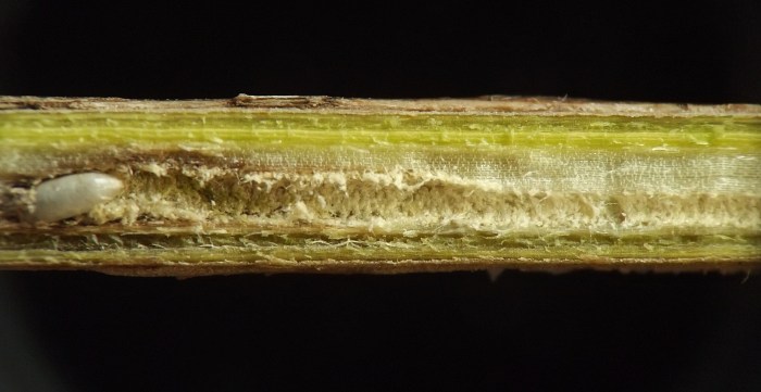
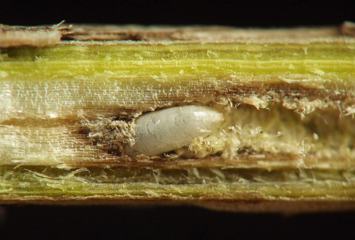
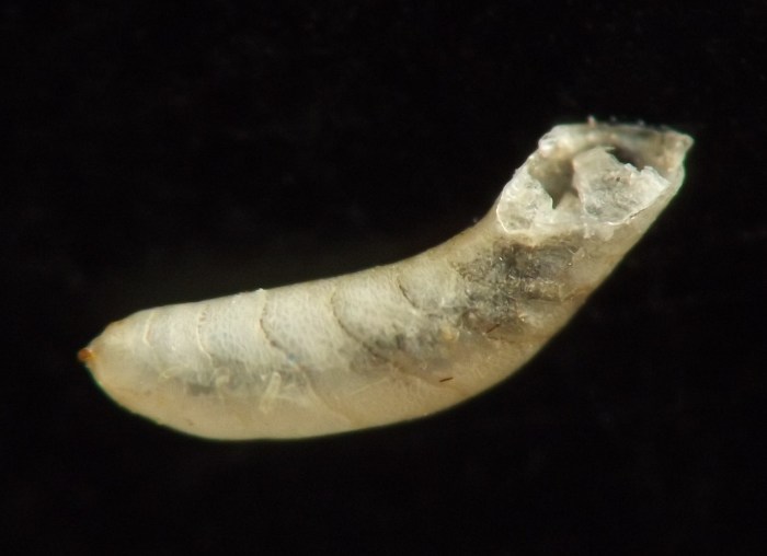
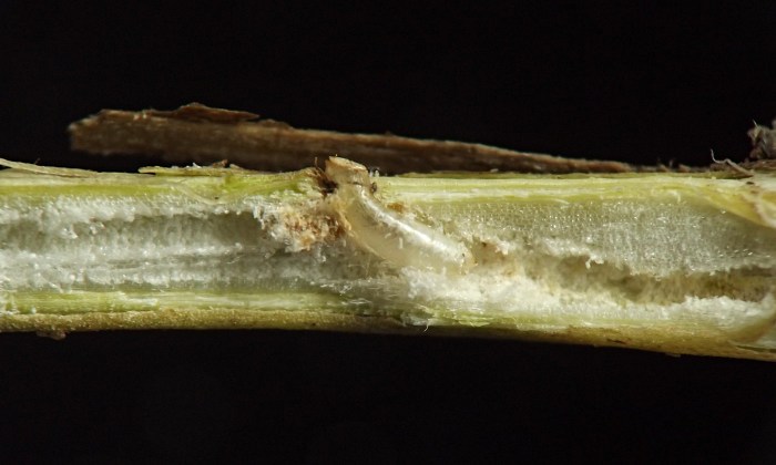
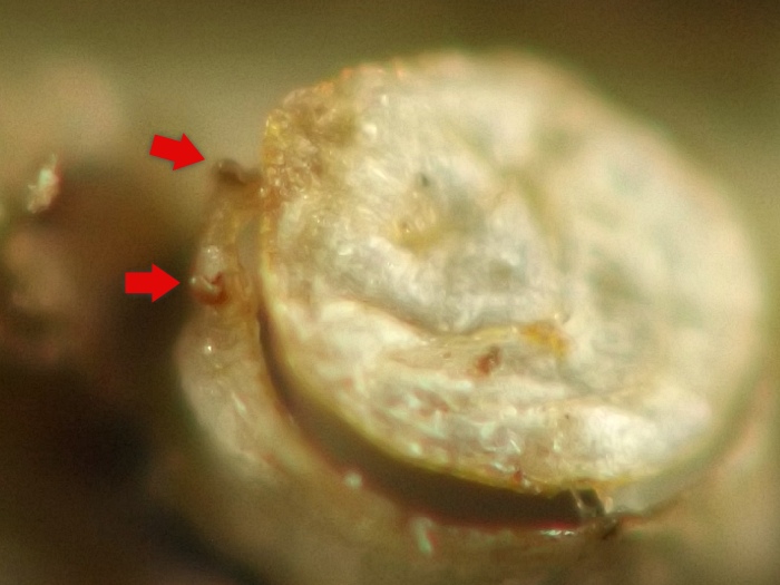
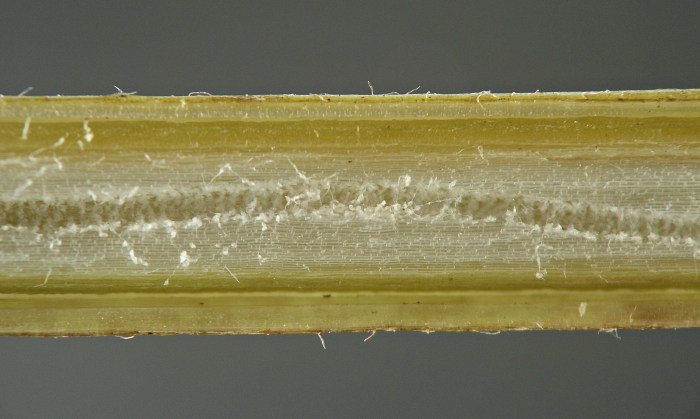
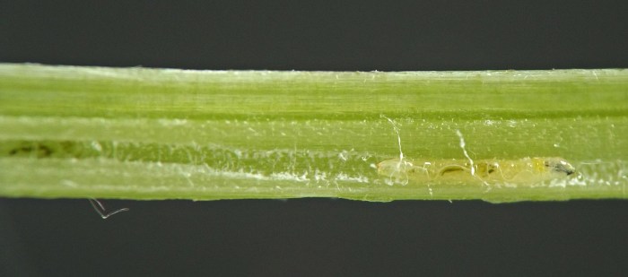
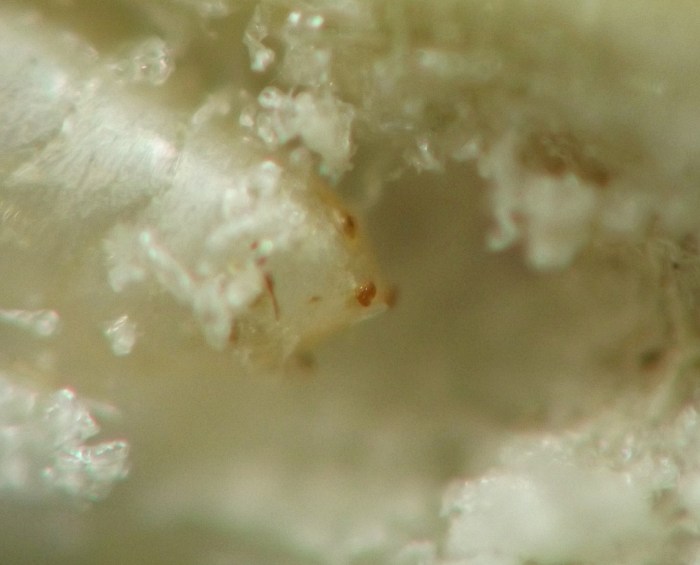
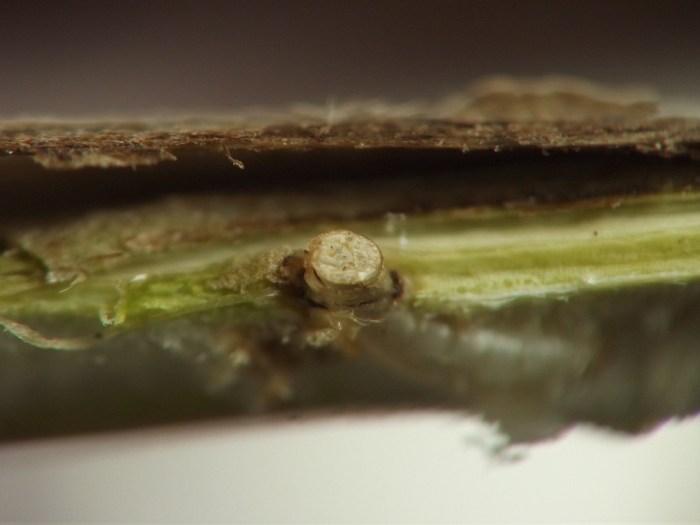
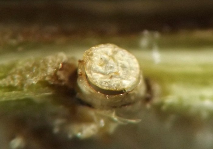
The larva of this borer forms an elongate tunnel in the pith of the stem. Rather than concentrating its feeding in a localized area within the stem, this species travels for a long distance along the main axis of the stem until it completes its development, its tunnel usually spanning multiple internodes. When it is finished feeding, usually somewhere along the length of an internode, it establishes a short, curving tunnel outward to just under the bark of the stem. It cuts an exit slit or window in the bark that remains closed for the time being. The larva then pupates in the curved tunnel, the puparium taking on the arching shape of the tunnel in a manner unusual among the stem agromyzids examined in the current study. Also unusual for a stem agromyzid, the anterior end of the puparium is modified into a peculiar, flat, circular "lid" that remains appressed to the underside of the bark until the adult emerges, at which point the "lid" (along with the exit window in the bark) pops open. The anterior spiracles of the puparium arise from the main body of the puparium, projecting from the rim where the "lid" attaches.

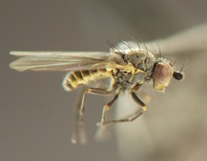
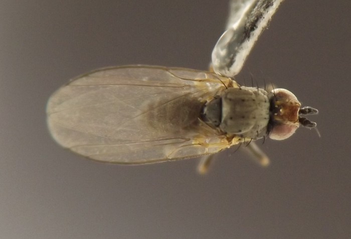
A single male adult was reared in spring 2023.
IMG#: 0159-21
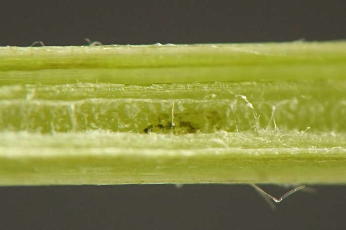
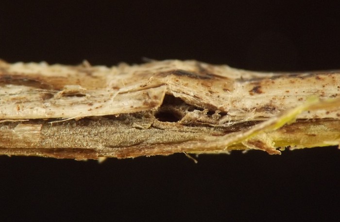
Stem of host, dissected, showing larva's tunnel.
IMG#: 0159-01
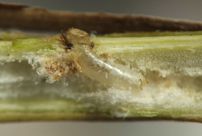
Another dissected stem, showing tunnel occupied by larva. (Photo date: July 13, 2022)
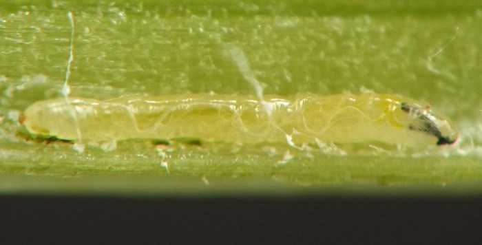
IMG#: 0159-02
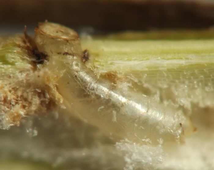
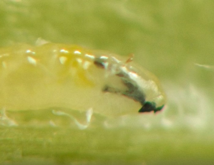
Larva in tunnel in stem. (Photo date: July 13, 2022)
IMG#: 0159-03
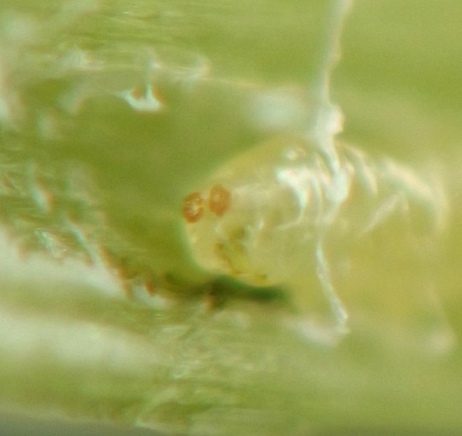
Lateral view, anterior end of larva. Note the shape of the anterior spiracles. Also note the way the posteriormost portion of the cephalopharyngeal skeleton appears divided in two (not three), indicating the larva belongs to subfamily Phytomyzinae.
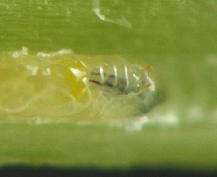
IMG#: 0159-04
Dorsolateral view, anterior end of larva.
IMG#: 0159-05
Posterior spiracles of larva.
IMG#: 0159-06
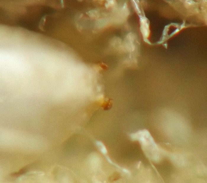
Detail, anterior end of larva in lateral view. The image, though mostly out of focus, is intended to give a sense for the appearance of the larva's mouthhooks. (Photo date: July 13, 2022)
IMG#: 0159-28
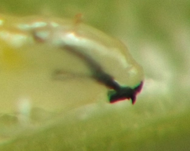
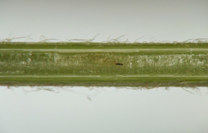
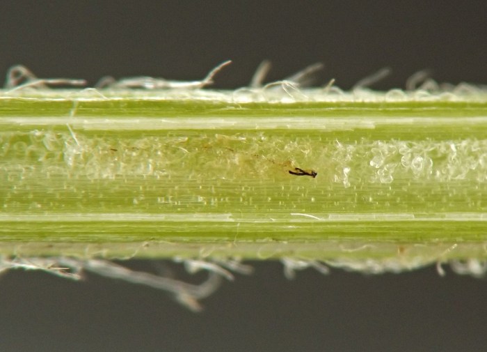
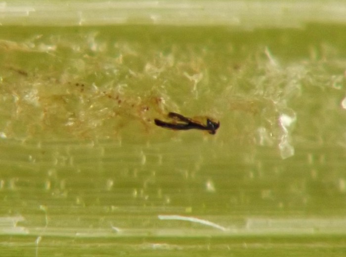
Remnants of a larva's cephalopharyngeal skeleton (black wishbone-shaped object), apparently resulting from a molt, left behind in the larva's tunnel in the stem. Note that the tunnel on both sides of the remains, and especially to the right, appears to be filling in with callus tissue. (Photo date: July 7, 2023)
IMG#: 0159-29
A closer view of the remnants of a larva's cephalopharyngeal skeleton, apparently resulting from a molt, left behind in the larva's tunnel in the stem. Note that the tunnel on both sides of the remains appears to be filling in with callus tissue.
IMG#: 0159-30
Remnants of larva's cephalopharyngeal skeleton in tunnel in stem.
IMG#: 0159-07
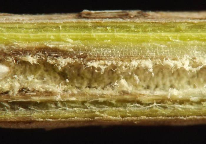
Larval frass in tunnel in stem. (Photo date: July 13, 2022)
IMG#: 0159-08
Endpoint of a larva's tunnel, with a spent puparium partly visible inside. (Photo date: May 3, 2021)
IMG#: 0159-09
A closer view, endpoint of a larva's tunnel, with a spent puparium partly visible inside. The anterior end of the puparium is out of view as it curves toward the far surface of the stem.
IMG#: 0159-10
Segment of completed tunnel.
IMG#: 0159-11
Spent puparium extracted from stem in early spring. Note unusual curved shape. The age of the stem was unrecorded, but it was likely a second- or third-year stem, with the adult having emerged from the puparium in a prior year when the stem was younger. (Photo date: May 3, 2021)
IMG#: 0159-12
Posterior spiracles of spent puparium.
IMG#: 0159-13
Hole in bark through which adult emerged.
IMG#: 0159-14
A second example of a spent puparium in a stem. The stem has been split open lengthwise for this view and the bark of the stem has been mostly removed. (Photo date: May 11, 2021)
IMG#: 0159-15
Spent puparium in stem.
IMG#: 0159-16
Spent puparium in stem.
IMG#: 0159-17
Posterior spiracles of spent puparium.
IMG#: 0159-18
More or less frontal view of anterior end of spent puparium in dissected stem.
IMG#: 0159-19
Detail, lidlike anterior end of spent puparium.
IMG#: 0159-20
Lidlike anterior end of spent puparium; the anterior spiracles, projecting from the main body of the puparium on the rim where the lid was attached, are indicated by the red arrows. (Photo date: May 11, 2021)
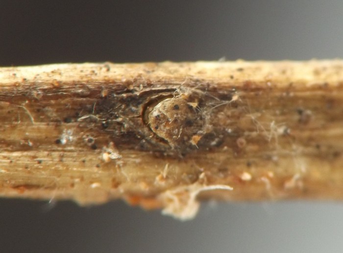
IMG#: 0159-22
Exterior view of affected stem at location where an adult recently emerged in springtime.
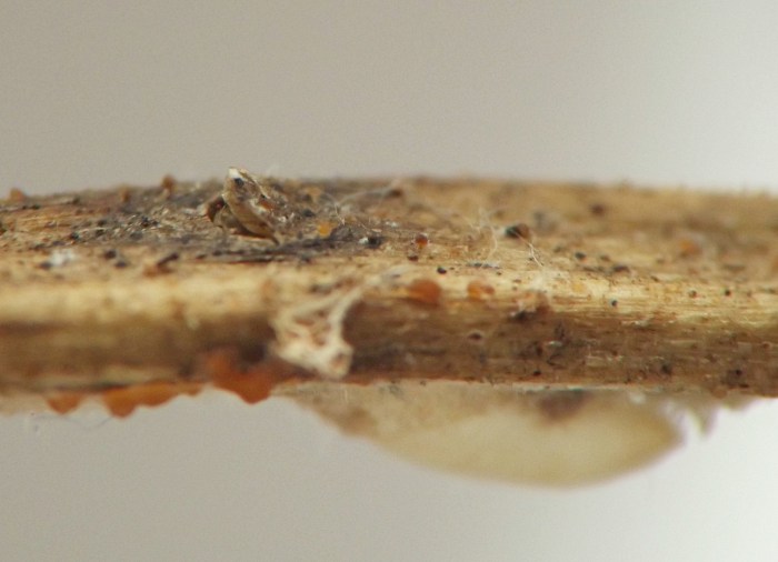
IMG#: 0159-23
Outer layers of tissue of a dissected stem, at the location of an adult fly's recent emergence, with the spent puparium still lodged in the tissue. The posterior part of the puparium is visible but out of focus below the brownish outer layers of stem tissue, and an exit window in the stem and the lidlike anterior end of the puparium have popped or hinged open to allow the adult to emerge.
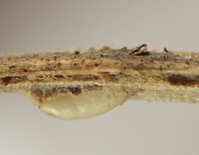
IMG#: 0159-24
Another view, outer layers of tissue of a dissected stem, at the location of an adult fly's recent emergence, with the spent puparium still lodged in the tissue. The posterior part of the puparium is visible but out of focus below the brownish outer layers of stem tissue, and an exit window in the stem and the lidlike anterior end of the puparium have popped or hinged open to allow the adult to emerge.
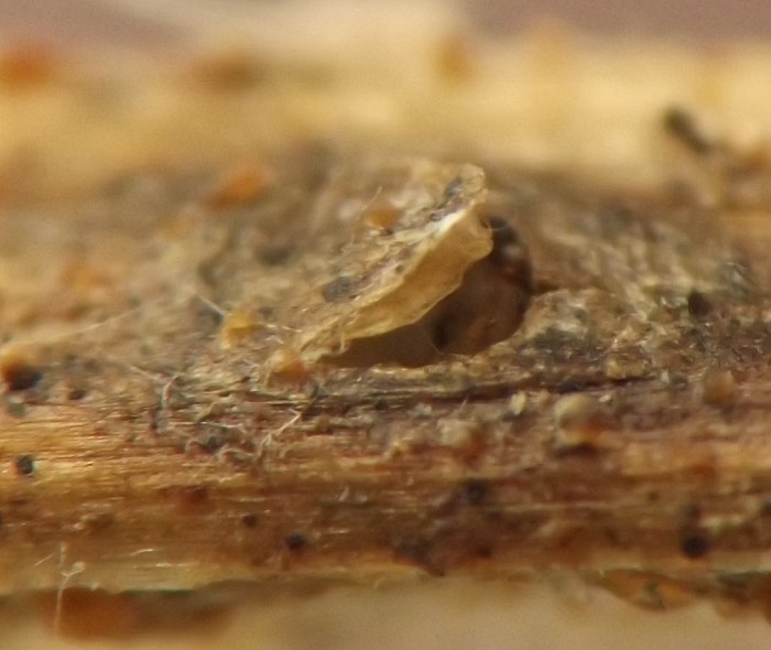
IMG#: 0159-25
The outermost layer of stem tissue and the lidlike anterior end of the puparium have both popped or hinged open to allow the adult fly to emerge. Barely discernable are the anterior spiracles of the puparium, out of focus and in shadow below the double "lid."
IMG#: 0159-26
Lateral view of adult fly reared from the puparium in the previous images (0159-22 through 0159-25). (coll. as puparium 01/05/23, adult em. 03/27/23)
IMG#: 0159-27
Dorsal view, reared adult.

References

[none]

Page created 10/16/23. Last update: 09/09/24