Local feeder (Diptera: Cecidomyiidae) in stems of Galium [0247]
| Order | Diptera |
|---|---|
| Family | Cecidomyiidae[T,L,P,A] |
| Lower taxon | undetermined |
| No. spp. involved | One confirmed (+1?) |
| Feeding mode | Local feeder in stem |
| Host plant | Cleavers, Galium aparine (Rubiaceae) |
Several Galium aparine whole plants (not including roots) were harvested from the author's yard in the final days of June, 2023, in an effort to rear an agromyzid that feeds internally in the stems of this plant. At the time of harvest, all leaves, flowers, and fruits were stripped from the plants, so that stems were the only plant material going into the rearing containers. The plants had mostly finished their growth and were undergoing senescence when they were harvested. The stems were divided up between three rearing bags.
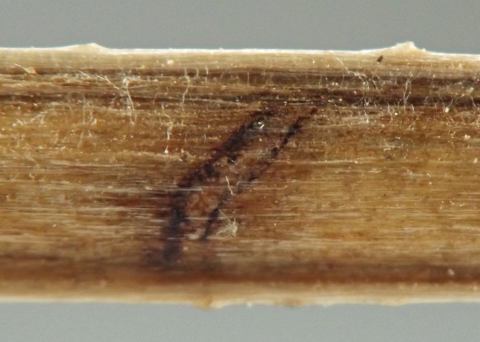
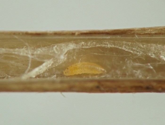
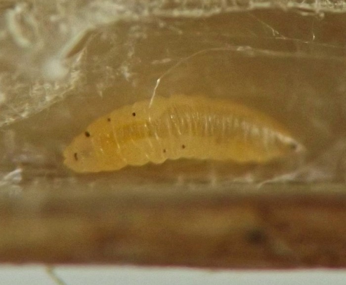
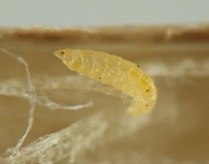
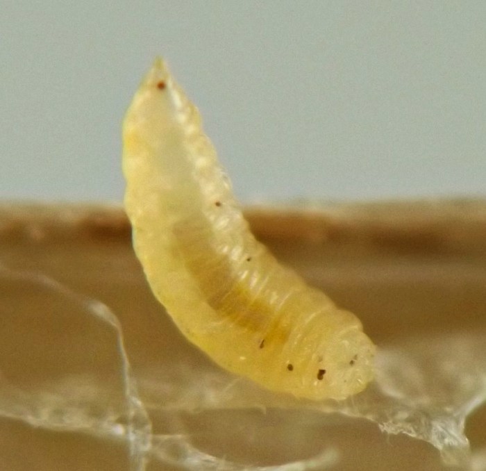
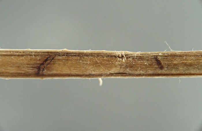
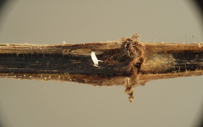
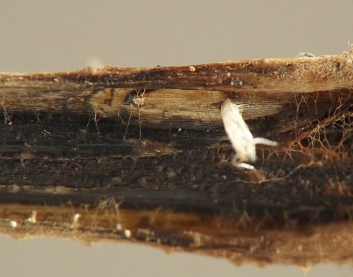
On 08 July some of the harvested stems were removed from one of the rearing bags and searched for agromyzid larvae. One of these stems had no agromyzid larvae inside but instead was found to possess a set of two externally visible, transverse dark brown or black marks, separated by a distance of approximately 10mm. Inside the stem between these two marks were two pale yellow-orange cecidomyiid larvae. It was unclear if the marks were related to the presence of the larvae. The larvae were transferred to another container and the stems remaining in the original three rearing bags were left undisturbed.
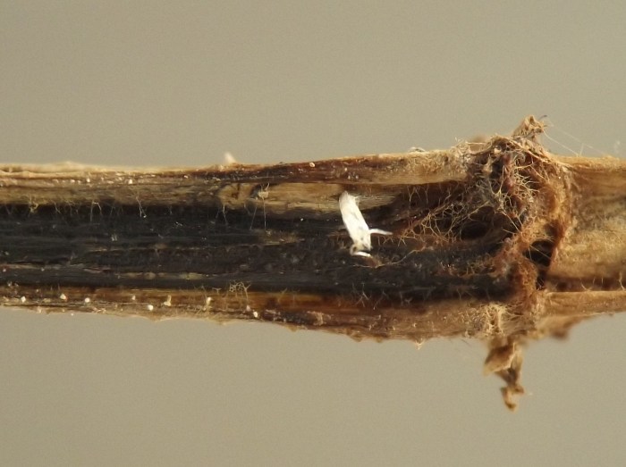
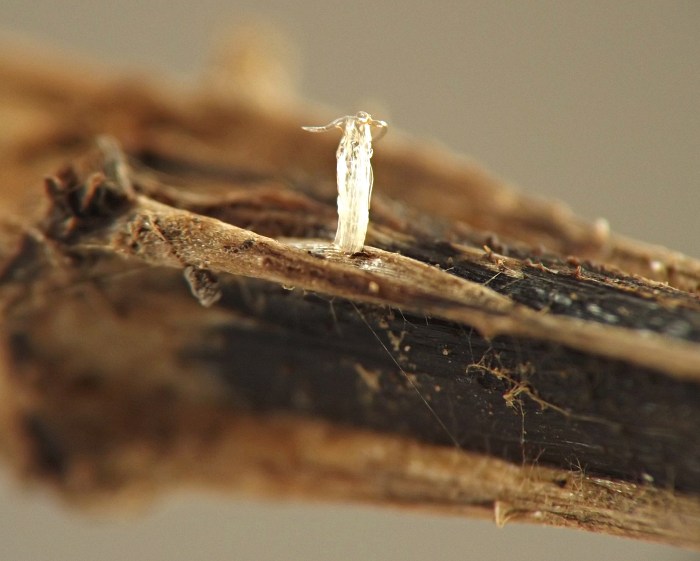
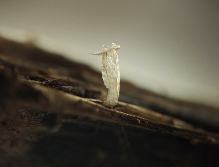
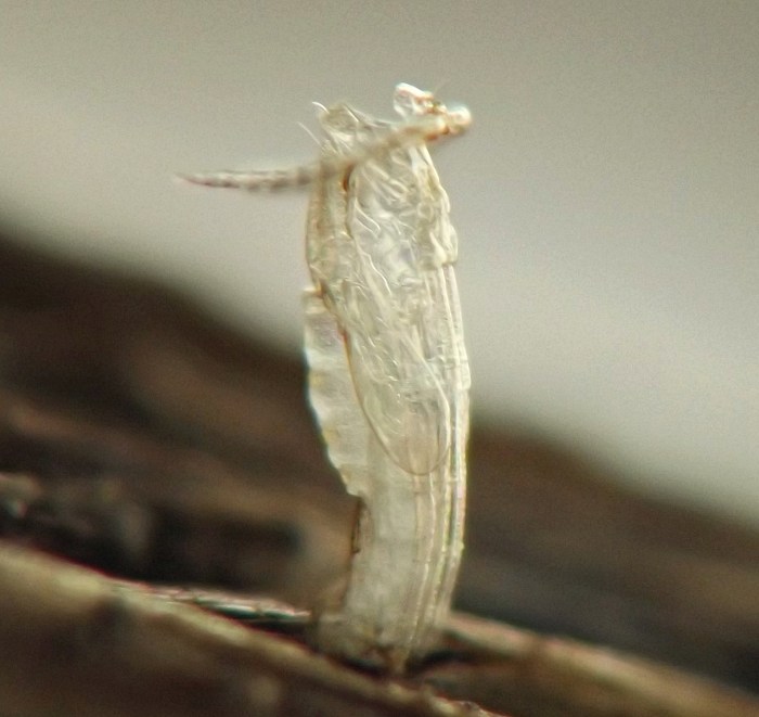
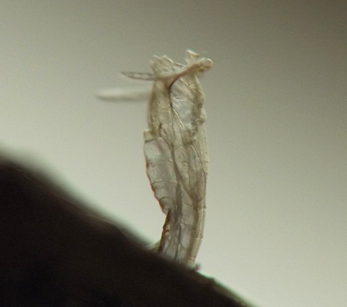
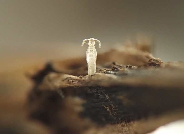
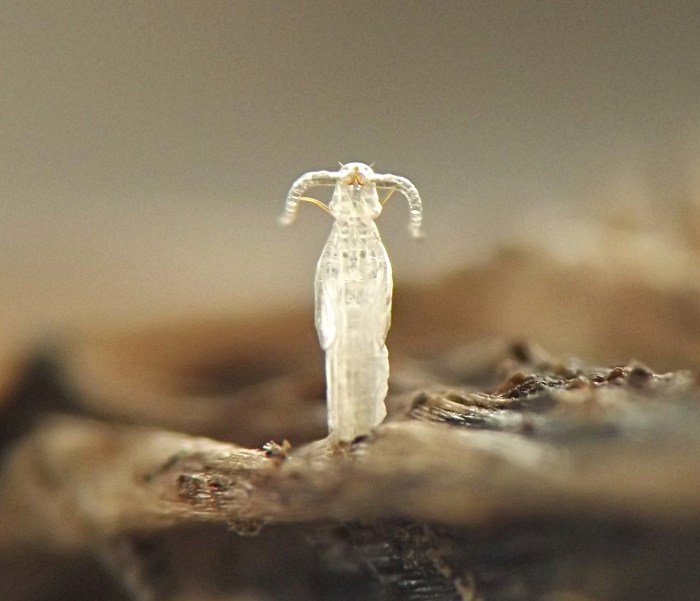
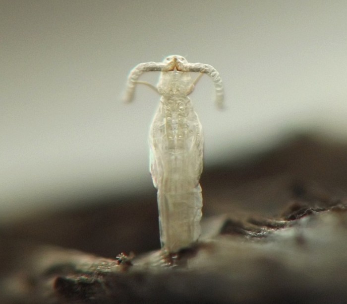
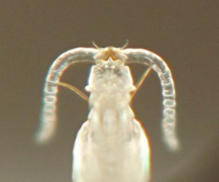
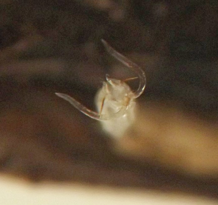
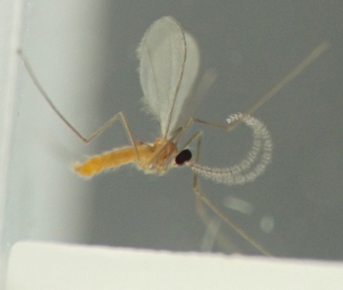
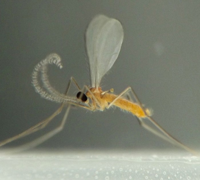
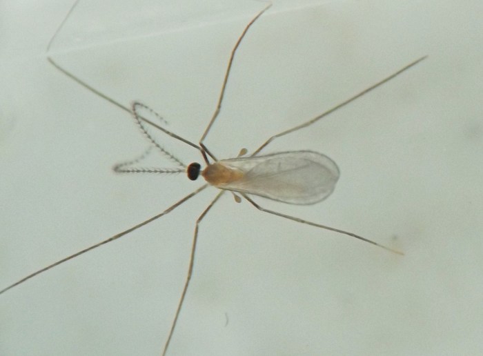
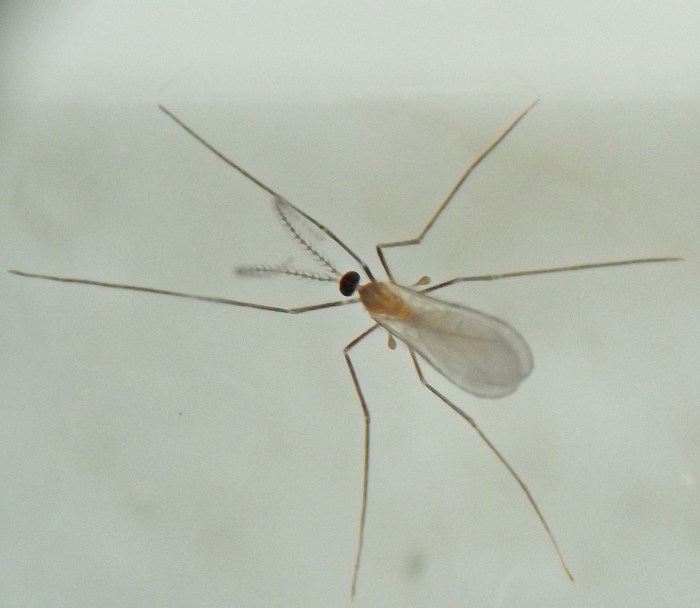
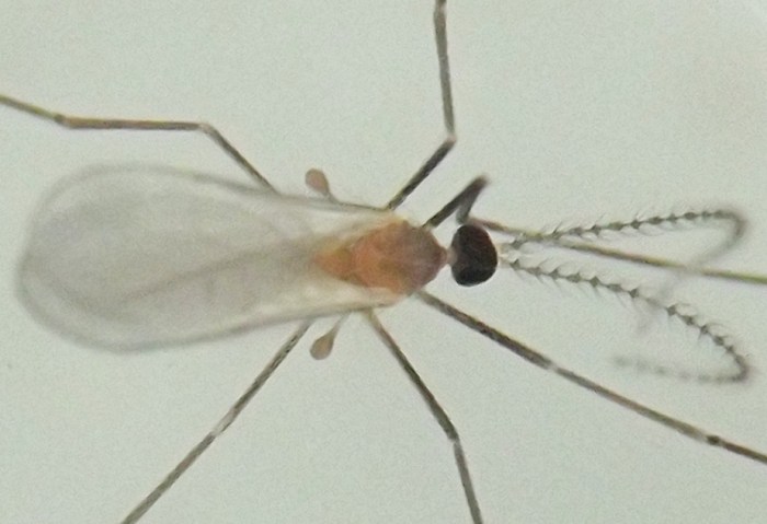
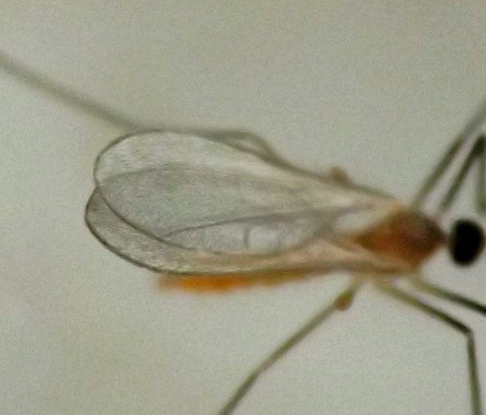
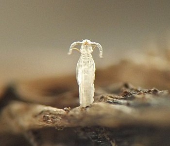
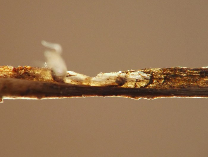
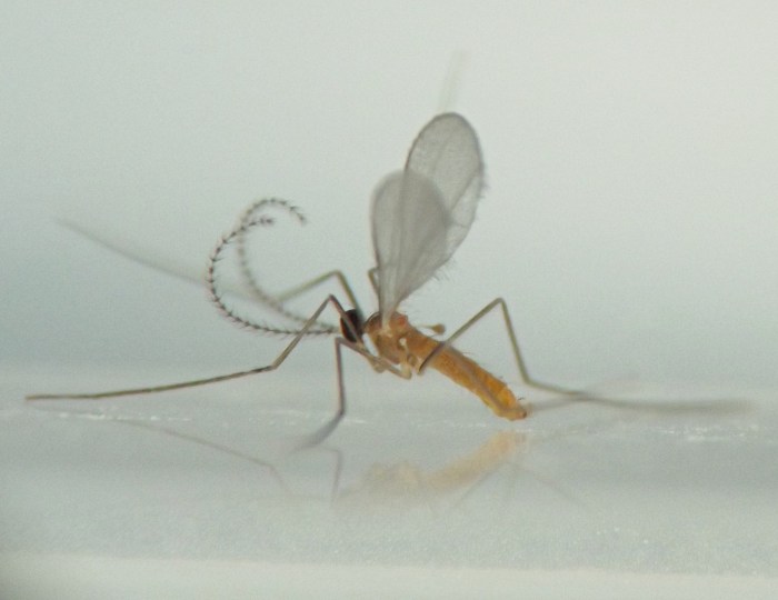
On 31 July a single adult cecidomyiid emerged in one of the rearing bags. After the adult had been collected from the bag, all the stems from this bag were searched carefully for protruding pupal exuviae. Eventually, with much effort, a single exuvium was found. It was protruding from a small, whitish, elongate-oval, slightly raised, blisterlike chamber just under the epidermis of the stem near a node. The exuvium was clearly from a cecidomyiid, and the initial assumption was that it belonged to the adult that had appeared in the bag, but this could not be immediately confirmed.
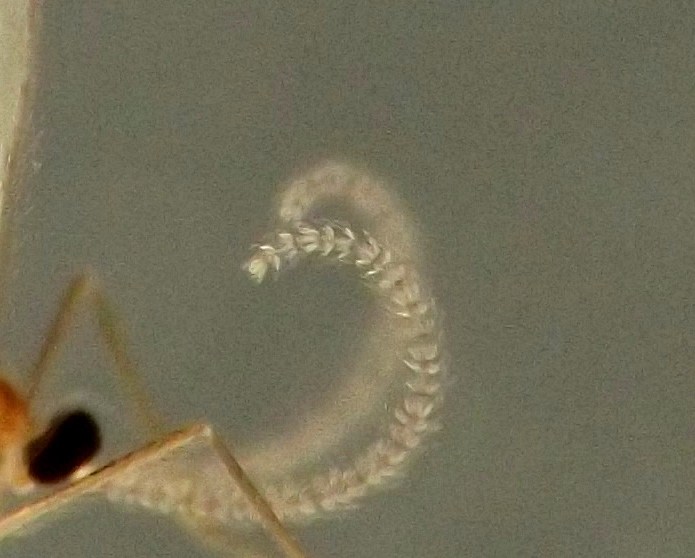
The larvae, pupal exuviae, and adult are shown in the photos below. Because the plant material had already begun to senesce when it was collected, it is conceivable that the stems could have been favorable habitat for a cecidomyiid feeding on fungal rather than plant material. However, the blisterlike chamber from which the exuviae protruded was suggestive of a gall formed by a plant feeder. Eventual identification of the specimens involved may shed light on the situation.
Specimen data for images
Coll. 06/27/23-06/28/23, larvae found in stem and photographed on 07/08/23 (01-06), adult and exuviae found in rearing container and photographed on 07/31/23 (07-27).
References
[none]
Page created 11/20/24. Last update: [none]


