Stem borer (Diptera: Agromyzidae) in Galium (#0244)[0244]

OrderDiptera
FamilyAgromyzidae[M,L,P,A]
Lower taxoncf. Ophiomyia
No. spp. involvedOne confirmed [1-C]
Feeding modeStem borer
Host plantBedstraw, Galium ?triflorum (Rubiaceae)
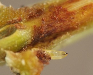
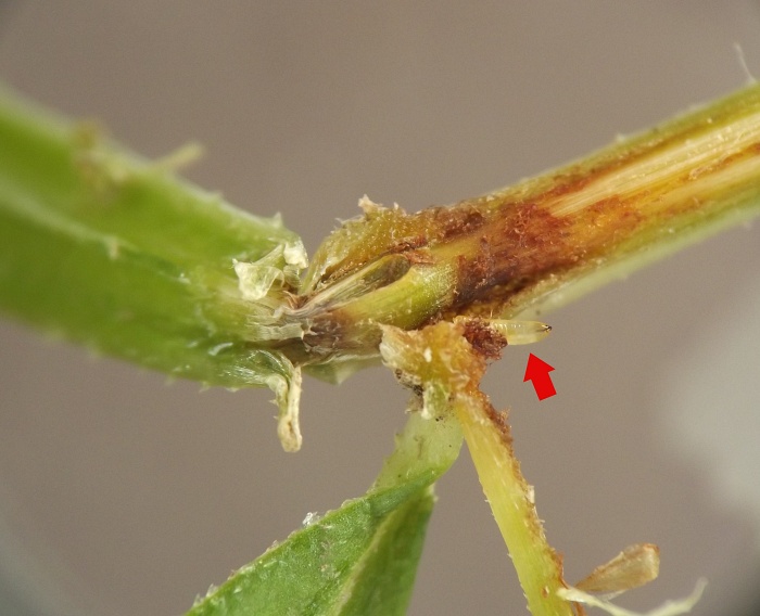
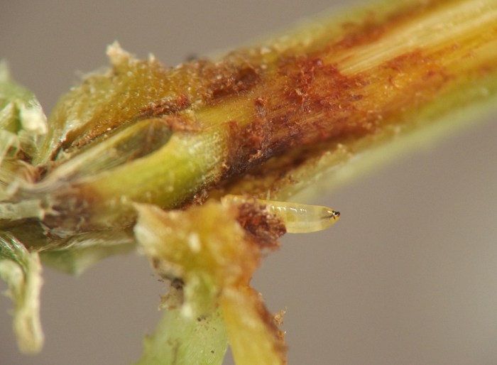
Young larva in between inner and outer layers of stem node

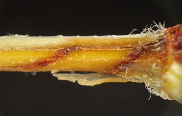
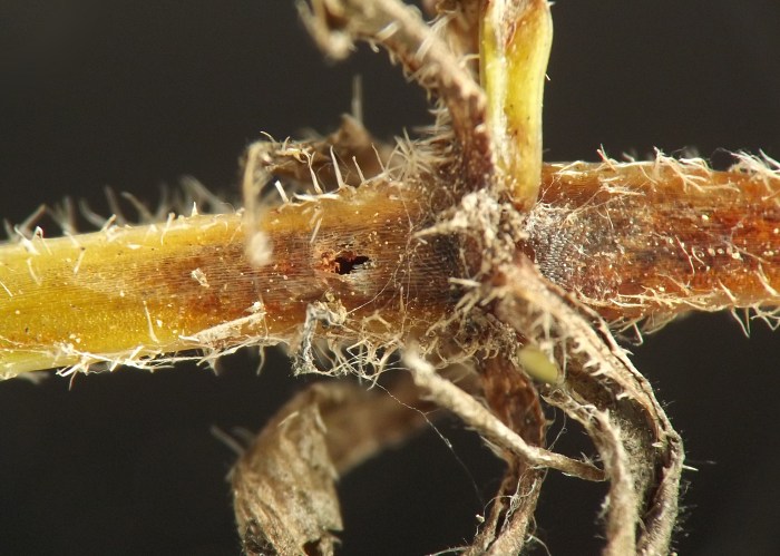
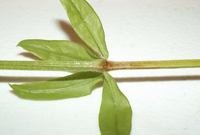
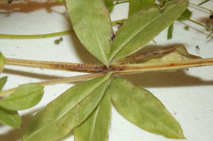
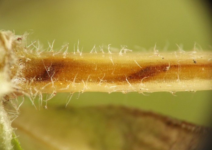
The externally visible sign of this borer consists of rusty reddish-brown discoloration to the stems, especially at the nodes but also in the form of shallow subsurface linear tracks along the internodes. Depth-wise, the larva's feeding mostly occurs in the space between the outer sheathlike layer and the tough inner core layer of the stem, and the outer layers of tissue are generally thin enough to allow the rusty discoloration to show through. The larva tunnels shallowly along the lengths of the internodes, with the tunnel sometimes spiralling around the stem but usually not doubling back on itself (i.e., an individual larva apparently only travels the length of a given internode once). Upon reaching a node, the larva performs some more concentrated excavation of the inner tissues, and if it is still young, after a little while it will leave the partially excavated node and enter the next internode to continue its movement and feeding along the length of the stem. After moving through multiple nodes and internodes in this way, the larva finishes its development in a node, which it more or less completely hollows out with the possible exception of some of the inner core layer. The larva then prepares an exit window in the epidermis and forms its black puparium just beneath the exit window within the hollowed-out node.

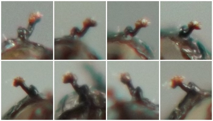
The anterior spiracles of the final-instar larva and puparium feature an archlike protrusion at the base of the spiracle stalk that projects slightly above the main body of the puparium, but not as prominently as the distal end of the stalk.

Adults were reared in spring 2022. The adult and puparium both resemble Ophiomyia, and the recently described O. galiodes has been reared from Galium plant material in spring (see Lonsdale 2021), but its feeding mode was unrecorded. The current borer should not be confused with the Ophiomyia stem miner that may also be found in Galium stems in the study area (record #0242). The miner creates a blotch mine in the internode just under the epidermis, causing the affected area to turn a silvery or light brown color.
IMG#: 0244-01
Affected stem, showing rusty discoloration at node. (Photo date: July 9, 2021)
full size
IMG#: 0244-09
Affected stem, showing rusty discoloration centered at node. (Photo date: August 13, 2021)
full size
IMG#: 0244-10
Stem of Galium ?triflorum adjacent to node, showing spiralling rusty-colored tunnel of larva visible through stem epidermis.
full size
IMG#: 0244-11
Stem of Galium ?triflorum adjacent to node, with outermost layer of stem tissue removed, revealing spiralling rusty-colored tunnel of larva.
full size
IMG#: 0244-02
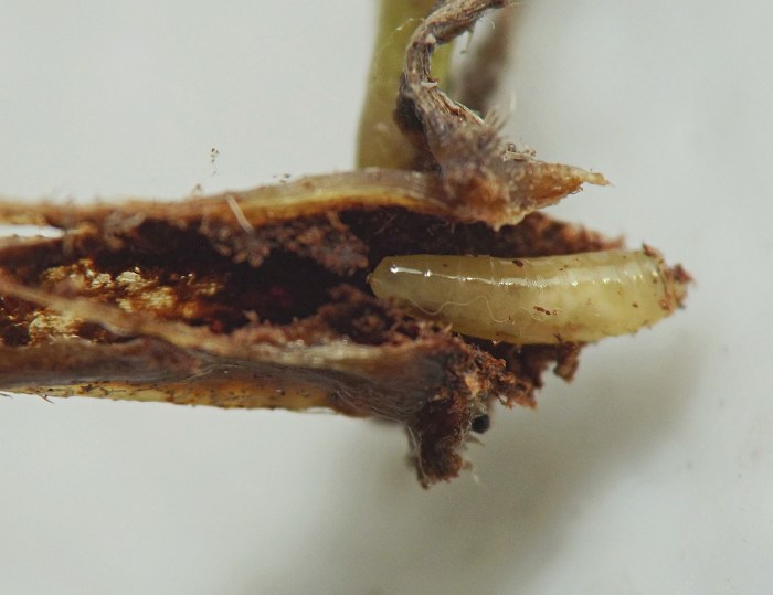
Young larva (arrow) in a dissected node. (Photo date: June 28, 2021)
full size
IMG#: 0244-03
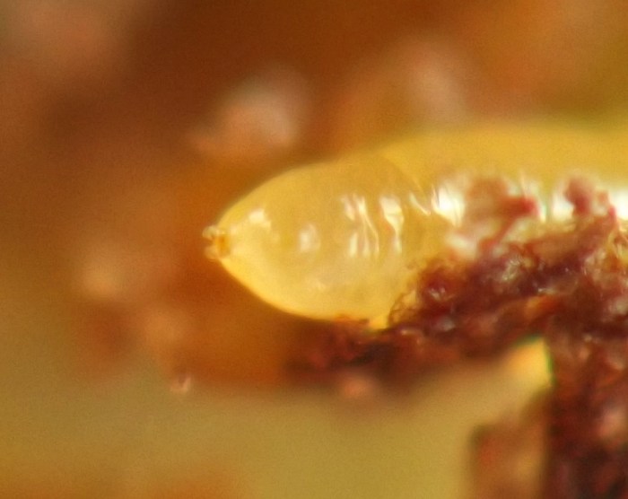
Young larva in a dissected node. (Photo date: June 28, 2021)
full size
IMG#: 0244-04
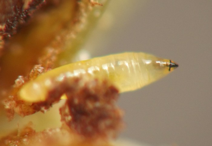
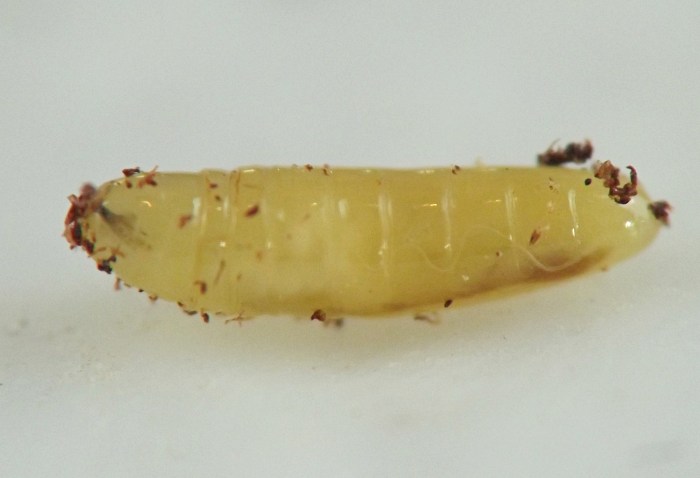
Ventral view of young larva from node of host.
full size
IMG#: 0244-05
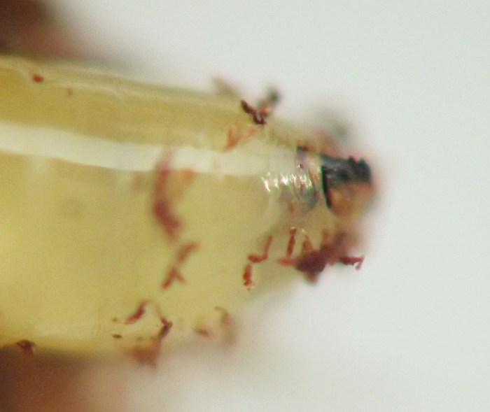
Ventral and ventrolateral views of young larva's anterior end.
full size
IMG#: 0244-06
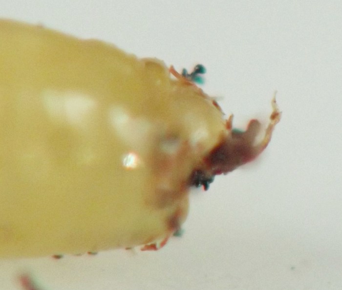
Posterior end of young larva.
full size
IMG#: 0244-07
Lateral view of young larva from node of host.
full size
IMG#: 0244-08
Another ventrolateral view of larva's anterior end.
full size
IMG#: 0244-12
Node inhabited by mature larva that has finished its feeding and mostly hollowed out the interior of the node. The small hole is part of the exit window the larva has established prior to pupating in the node. (Photo date: August 13, 2021)
full size
IMG#: 0244-13
Mature larva in hollowed-out node. (Photo date: August 13, 2021)
full size
IMG#: 0244-14
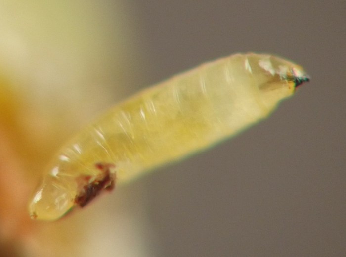
Mature larva, removed from node.
full size
IMG#: 0244-15
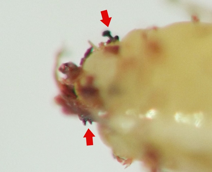
Anterior end of mature larva in lateral view, showing anterior spiracles (top arrow) and mouthhooks (bottom arrow).
full size
IMG#: 0244-16
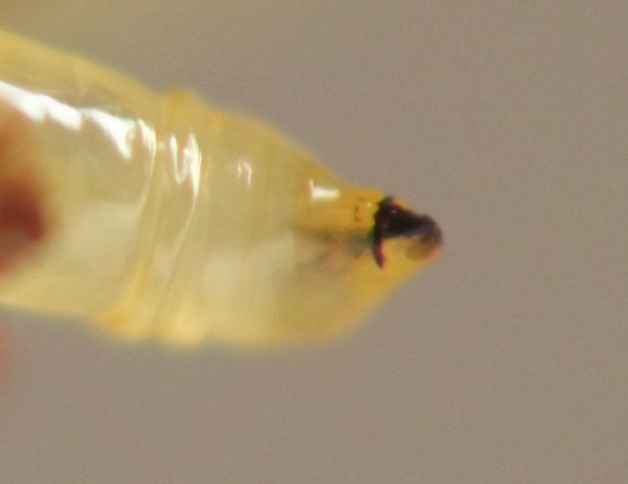
Anterior end of mature larva in lateral view.
full size
IMG#: 0244-17
Anterior end of mature larva in ventrolateral view.
full size
IMG#: 0244-31
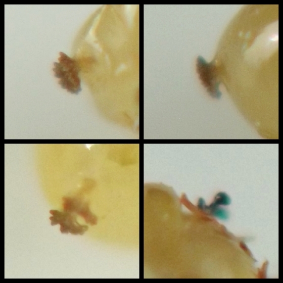
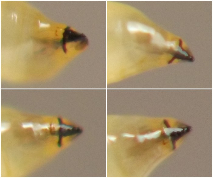
Views of a mature larva's anterior spiracles (bottom right panel) and posterior spiracles (other three panels).
full size
IMG#: 0244-18
Puparium, freshly formed. (Photo date: August 14, 2021)
full size
IMG#: 0244-19
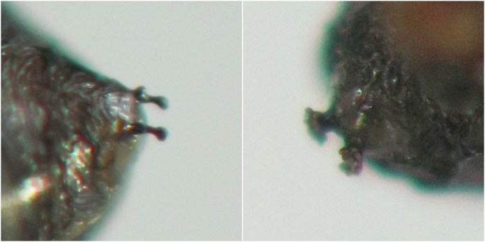
Anterior (left panel) and posterior (right panel) spiracles of freshly formed puparium.
full size
IMG#: 0244-34
Anterior spiracles of freshly formed puparium. Note the archlike protrusions at the bases of the spiracle stalks; see next image (0244-33) for better views of this feature on another puparium.
full size
IMG#: 0244-33
Several views of single anterior spiracles from a spent puparium, showing the archlike protrusion at the base of the spiracle stalk.
full size
IMG#: 0244-20
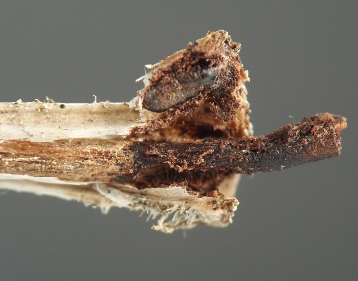
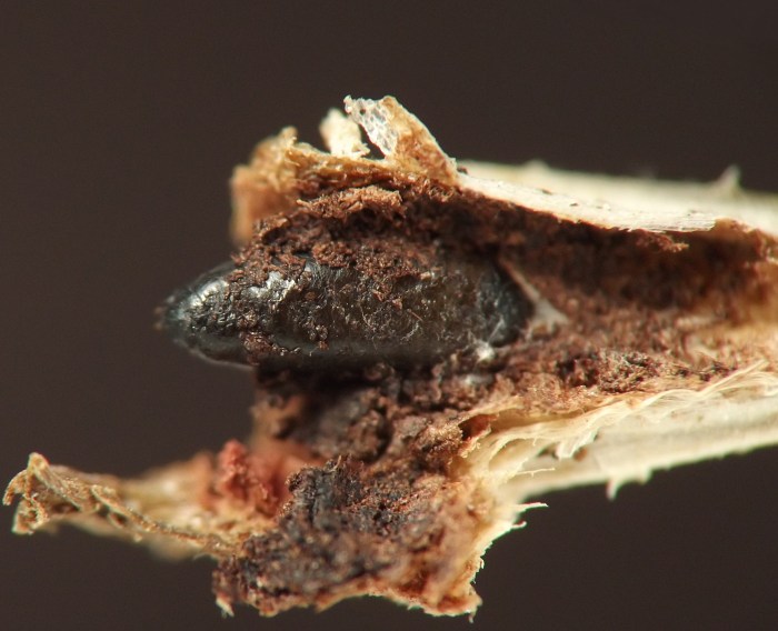
Overwintered dead stem of the host, dissected at the node where a larva finished its feeding and pupated in the prior growing season. The tough inner cylinder of plant tissue is all that remains of the inner tissue of the node; the rest has been eaten away. Note that even the outermost layers of the tough inner cylinder were scraped away by the larva, and the remaining tissue blackened as a result. The black puparium of the culprit is out of focus, above center. (Photo date: February 11, 2022)
full size
IMG#: 0244-21
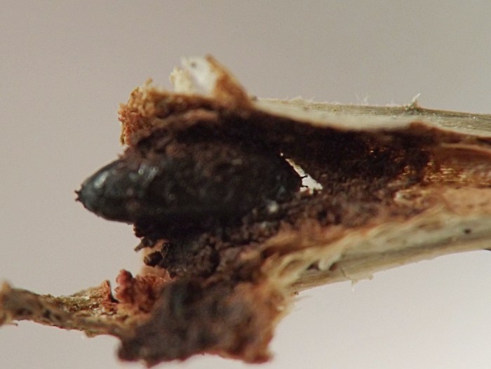
Puparium in hollowed-out node of overwintered dead stem of the host. (Photo date: April 4, 2022)
full size
IMG#: 0244-22
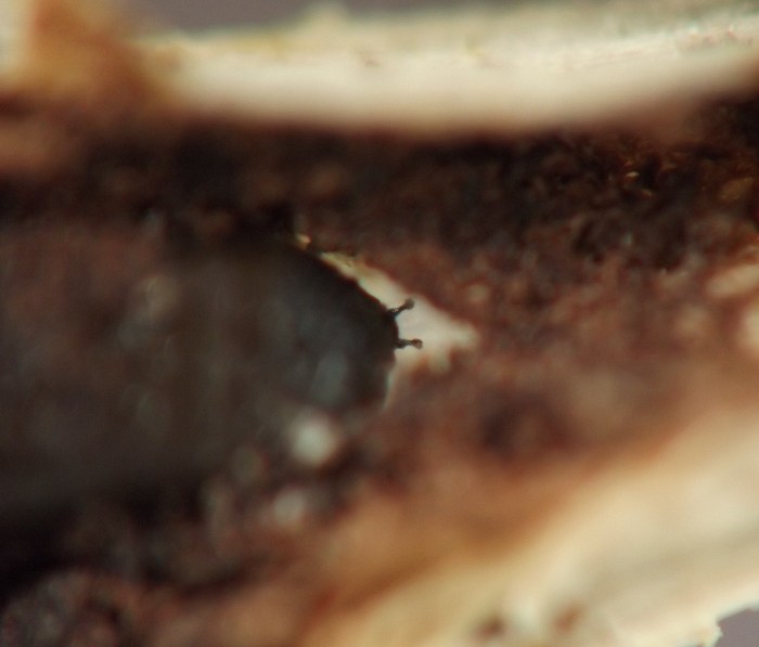
Another view of a puparium in a hollowed-out node of an overwintered dead stem, with this view showing the anterior spiracles in silhouette against the exit window cut by the larva prior to pupating.
full size
IMG#: 0244-23
Anterior spiracles of puparium, in silhouette against the exit window cut by the larva prior to pupating.
full size
IMG#: 0244-24
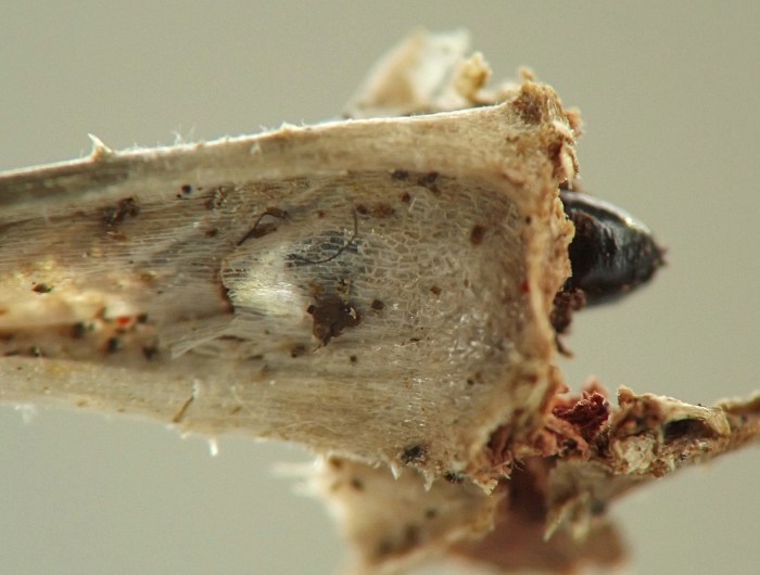
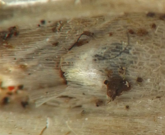
This photo shows the same specimen as the previous photo (0244-23), but from the opposite side, a view of the exterior of the inhabited node. The posterior end of the black puparium is visible to the right of center. Just to the left of center, the exit window for the adult-to-be is a lighter shade of gray than the surrounding tissue, a result of light being transmitted through it in this partially backlit view.
full size
IMG#: 0244-25
Detail of exit window from previous photo (0244-24).
full size
IMG#: 0244-26
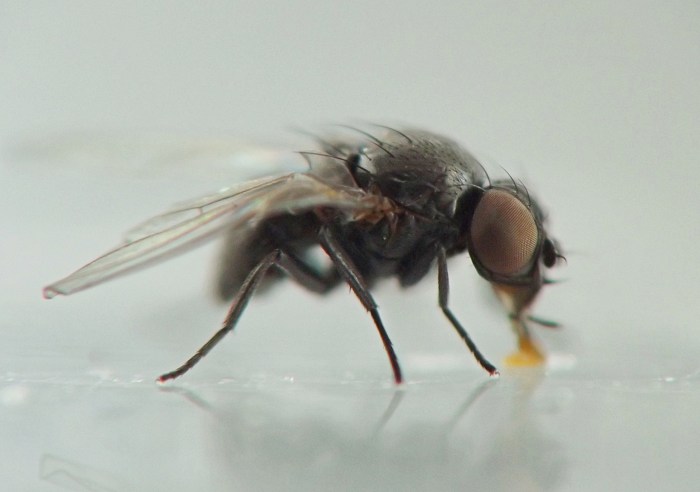
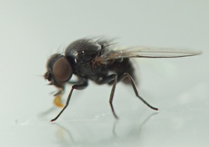
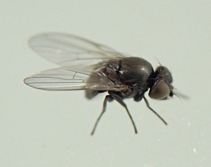
Reared adult, lateral.
full size
IMG#: 0244-27
Reared adult, fronto-lateral view.
full size
IMG#: 0244-28
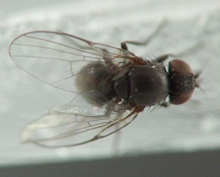
Reared adult, dorsal.
full size
IMG#: 0244-29
Reared adult, dorsolateral.
full size
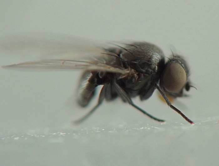
IMG#: 0244-30
Reared adult, lateral.
full size
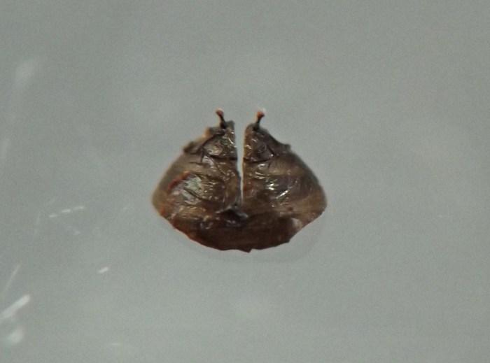
IMG#: 0244-35
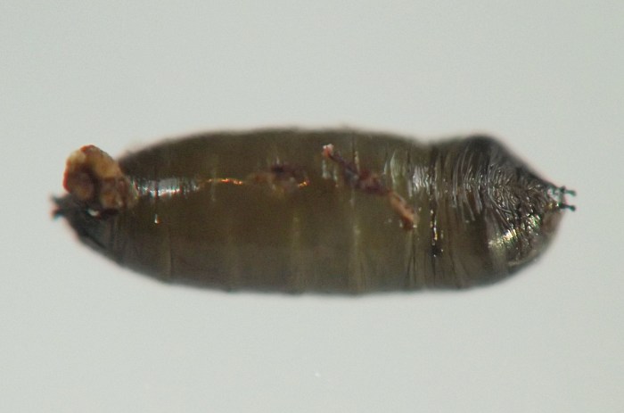
Dorsal-anterior portion of puparium, which split off from the rest of the puparium upon the successful emergence of the adult fly.
full size

Specimen data for images

Coll. 07/09/21, photographed same day (01); coll. 06/28/21, photographed same day (02-08); coll. 08/11/21, damage and larva photographed 08/13/21 (09-17, 31) and larva's fresh puparium photographed on 08/14/21 (18-19, 34); coll. 02/11/22, photographed same day (20); coll. 04/02/22, photographed 04/04/22 (21-25); coll. 02/11/22, em. 03/15/22 (26-28); coll. 04/02/22, em. 04/14/22 (29-30); coll. 04/02/22, adult em. by 04/16/22, puparium photographed on 10/27/23 (33, 35).

References

Lonsdale, O. 2021. Manual of North American Agromyzidae (Diptera, Schizophora), with revision of the fauna of the “Delmarva” states. ZooKeys 1051: 1–481. https://doi.org/10.3897/zookeys.1051.64603

Page created 10/27/23. Last update: 11/17/24