Stem borer (Diptera: Agromyzidae) in Parthenocissus [0359]

OrderDiptera
FamilyAgromyzidae[M,L,P,A]
Lower taxonundetermined
No. spp. involvedOne confirmed [1-C]
Feeding modeStem borer
Host plantParthenocissus sp. (Vitaceae)
Larva in stem of Parthenocissus

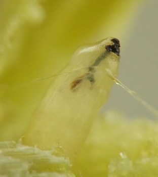
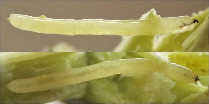
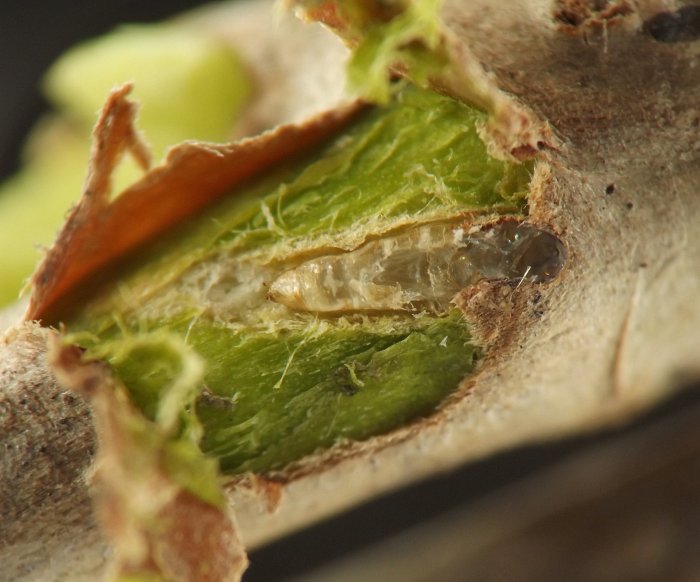
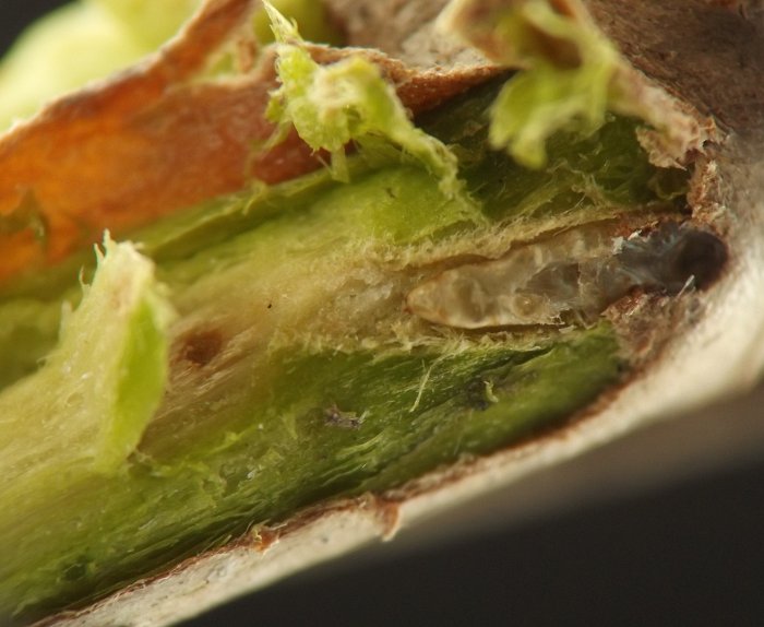
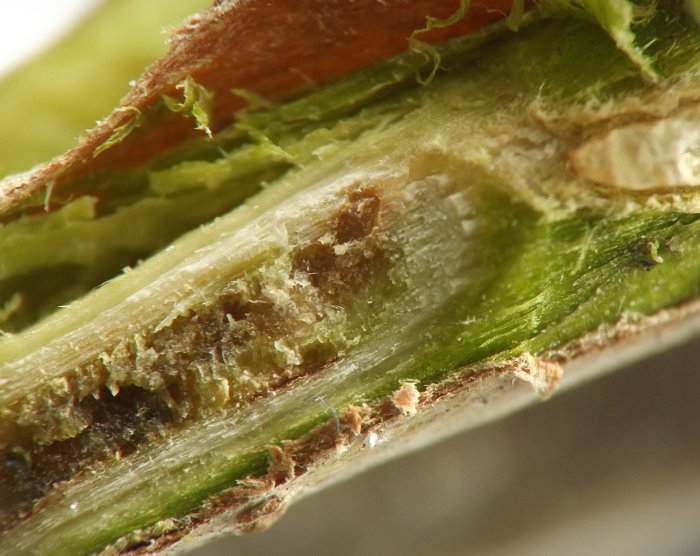
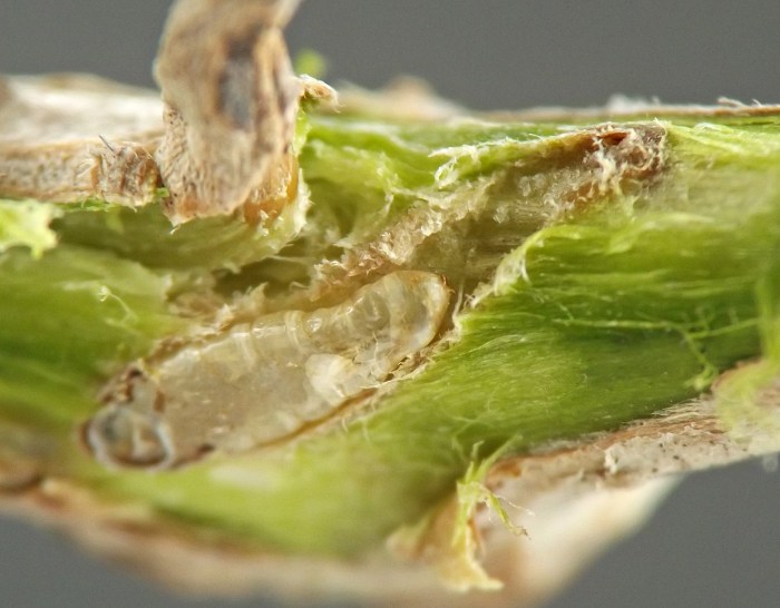
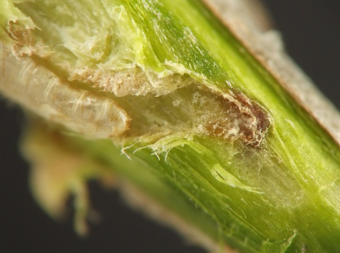
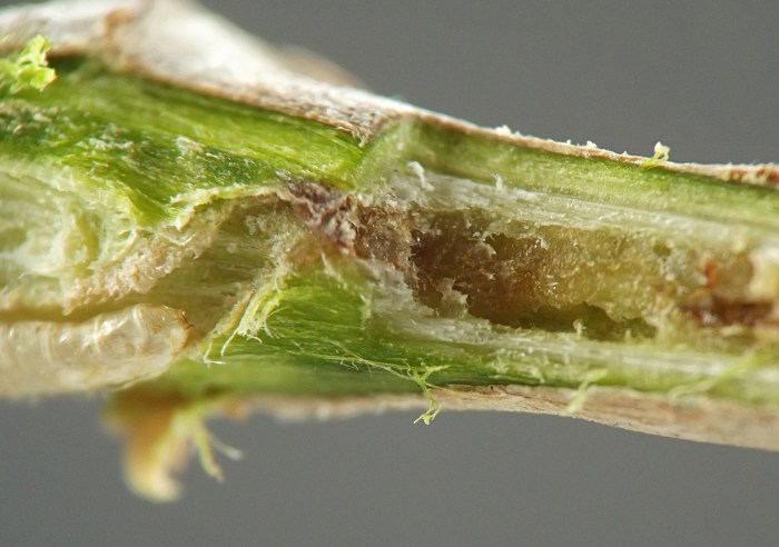
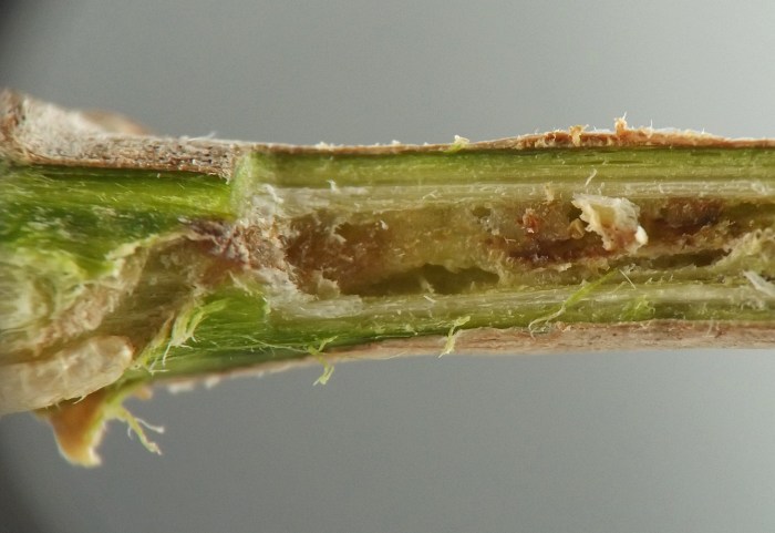
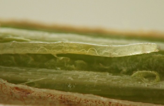
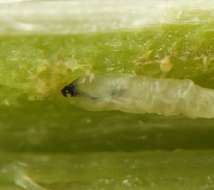
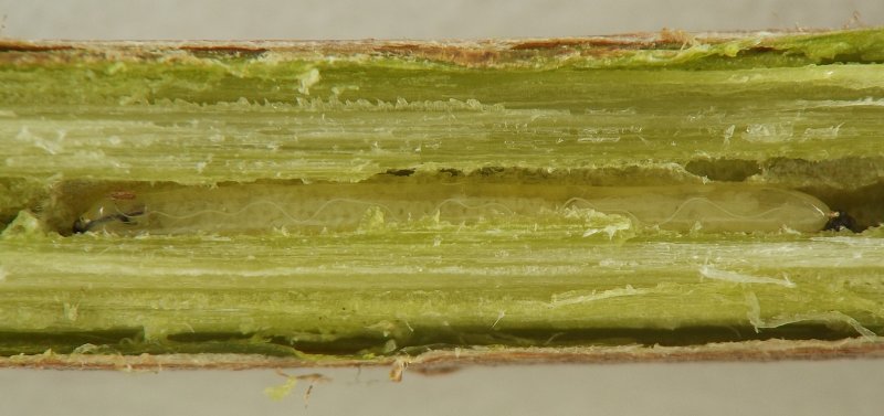
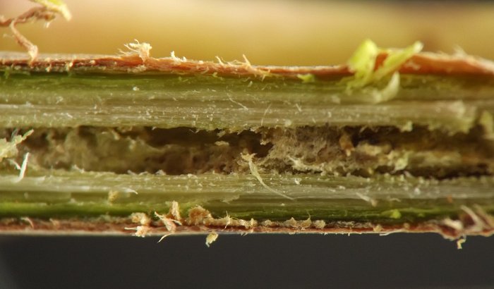
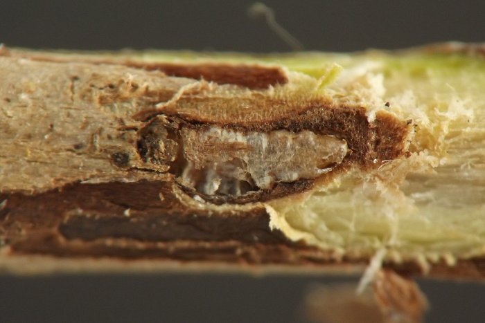
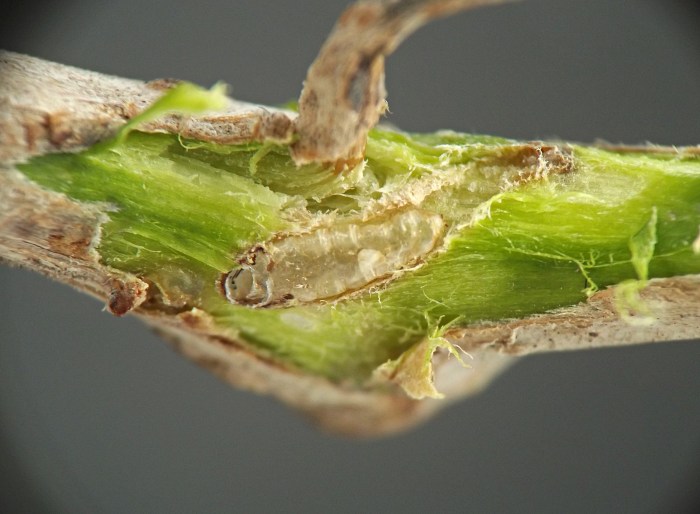
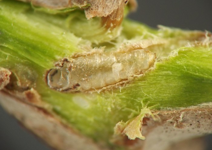
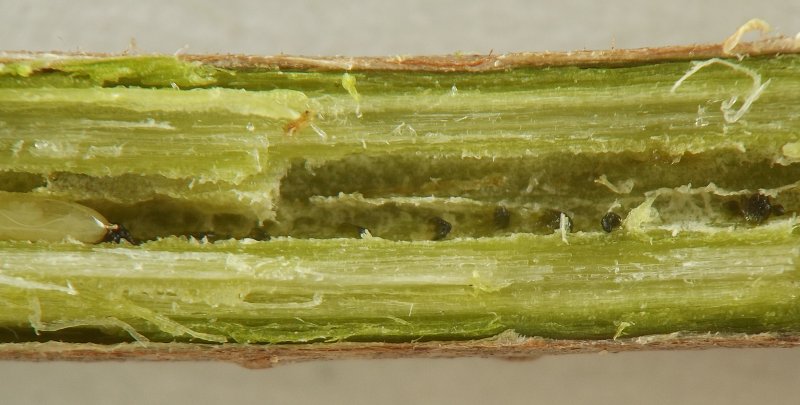
Larvae of this borer may initially be found tunneling in core tissues of young stems in summer. They are exceptionally long and thin. Their feeding creates irregular hollowing and disturbance to the stem interior, with at least some affected areas in the interior apparently forming callus tissue in response, as has been reported for tree tissues affected by Phytobia cambii in Europe (Ellis 2023). Dark greenish or blackish spots of frass are periodically distributed through the larval tunnels. At least some older larvae overwinter in the young woody stems; a few such overwintering larvae were located and photographed as part of the current study (see images below). Mature larvae migrate from deep in the stem interior to shallower tissues in order to pupate, creating an abbreviated (~10 mm) tunnel just under the bark, at the end of which the puparium is formed.

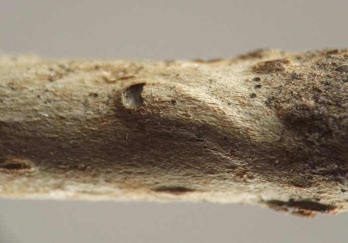
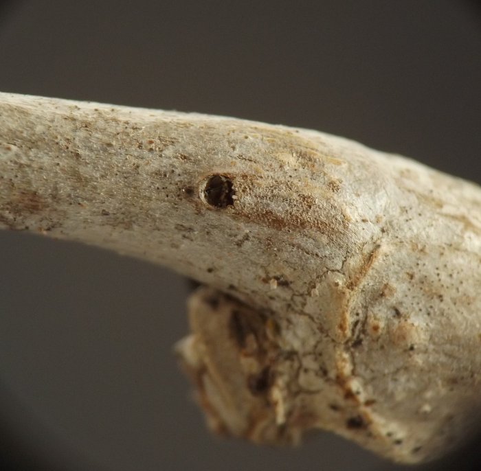
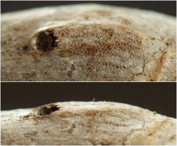
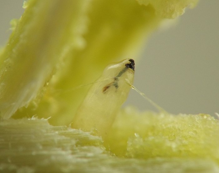
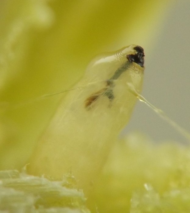
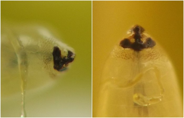
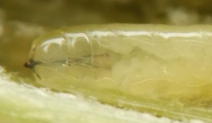
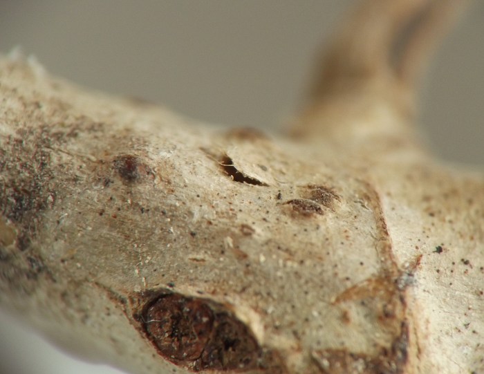
When the adult emerges, the anterior end of the puparium splits and, because this part of the puparium makes contact with the underside of the bark of the stem, a semicircular hole or exit slit in the bark (presumably cut by the larva prior to pupation) opens up simultaneously, through which the adult escapes. This is in contrast to most internally-pupating stem borer agromyzids the author has observed, for which there is usually a short but noticeable exit tunnel in the stem between the anterior end of the puparium and the operculum in the stem epidermis. The situation with no exit tunnel and in which the outermost layer of stem tissue opens up simultaneously with the anterior end of the puparium is more similar to what occurs with Ophiomyia spp. agromyzid stem miners who pupate just under the epidermis of herbaceous stems. However, unlike such stem miners, this Parthenocissus borer agromyzid either does not project its anterior spiracles through the outermost layers of stem tissue, or the anterior spiracles are subtle enough when so projected that the author did not notice them. The former scenario is favored given that the anterior spiracles of the larva seem to be somewhat reduced / recessed and not clearly stalked (see, e.g., image 0359-10 below).
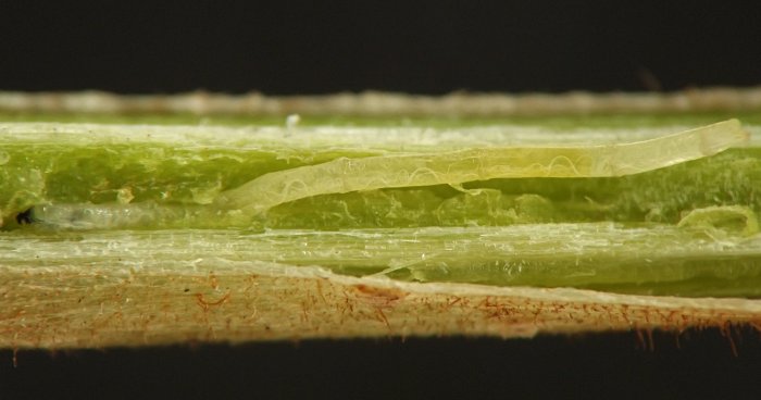
IMG#: 0359-01
Early- to middle-instar larva in young stem of Parthenocissus sp. in summer. (Photo date: August 19, 2018)
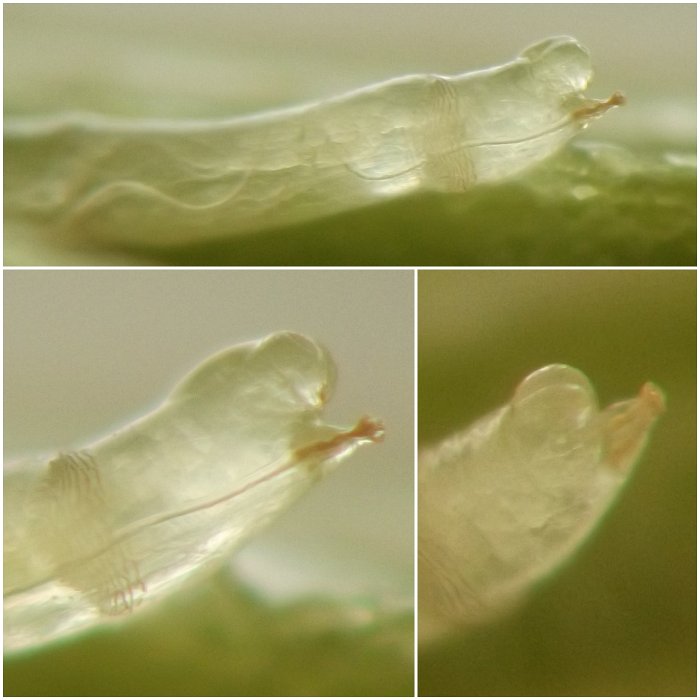
IMG#: 0359-02
Posterior end of early- to middle-instar larva.
IMG#: 0359-03
Anterior end of early- to middle-instar larva.
IMG#: 0359-04
Detail, posterior end of early- to middle-instar larva.
IMG#: 0359-05
Older larva overwintering in tunnel in stem interior. (Photo date: February 20, 2018)
IMG#: 0359-06
Area of tunnel immediately behind larva from previous photo (0359-05), showing several spots of dark greenish frass.
IMG#: 0359-07
Anterior end of older larva overwintering in stem. (Photo date: February 17, 2018)
IMG#: 0359-08
Detail, anterior end of older larva overwintering in stem.
IMG#: 0359-09
Lateral and ventral views of sclerotized areas on older larva's front end.
IMG#: 0359-10
Dorsal view, anterior end of older larva overwintering in stem.
IMG#: 0359-11
Posterior end of older larva, showing spiracles.
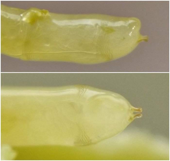
IMG#: 0359-12
Older larva, extricated from stem in winter; lateral (top panel) and ventral (bottom panel) views.
IMG#: 0359-40
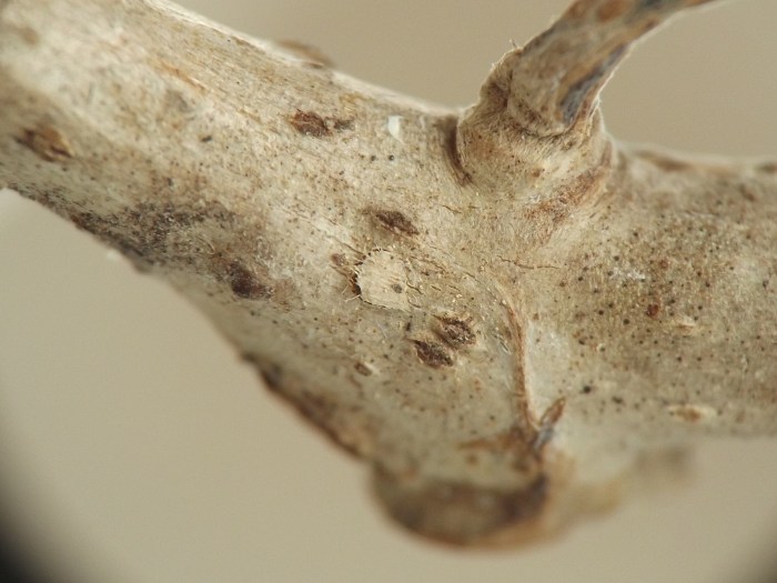
Bump on surface of stem, corresponding to location of spent puparium under bark, with a hole where the adult agromyzid emerged from the anterior end of the puparium. This example was found "as is" in midwinter, indicating the adult had emerged from the woody stem in a prior year. (Photo date: February 2, 2018)
IMG#: 0359-13
A second example of a bump on the surface of a Parthenocissus stem, corresponding to location of spent puparium under bark, with a hole where the adult agromyzid emerged from the anterior end of the puparium. This example was found "as is" in early April, indicating the adult had emerged from the woody stem in a prior year. (Photo date: April 9, 2022)
IMG#: 0359-14
Bump on stem surface, revealing the location of a spent puparium underneath, with adult's emergence hole in bark.
IMG#: 0359-29
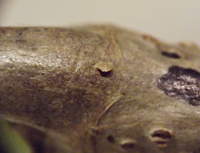
Another example of an adult agromyzid's emergence site, revealed by a semicircular exit flap in bark of stem.
IMG#: 0359-30
Semicircular exit flap of emerged adult in bark of stem.
IMG#: 0359-31
Semicircular exit flap of emerged adult in bark of stem.
IMG#: 0359-28
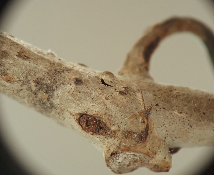
A third example of an adult agromyzid's emergence site. The emergence site was circled with a permanent marker to help the author keep track of which puparia were still intact and which ones were already spent on the day the stem was collected.
IMG#: 0359-16
A fourth example of an adult agromyzid's emergence site. Upon collecting this stem in April 2022, the author externally examined it for possible locations of intact puparia, and marked these by inscribing a black dot on the bark on either side of each location with a permanent marker. After a few weeks held in a rearing container indoors at room temperature, an adult emerged from a puparium at this location on May 4, leaving a semicircular exit slit in the bark. (Photo date: May 4, 2022)
IMG#: 0359-17
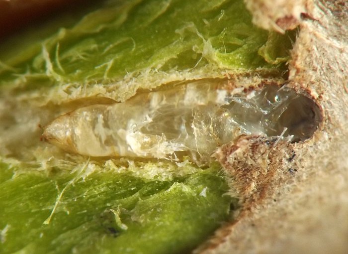
Location from previous photo (0359-16), dissected, revealing spent puparium under bark.
IMG#: 0359-18
Spent puparium under bark.
IMG#: 0359-19
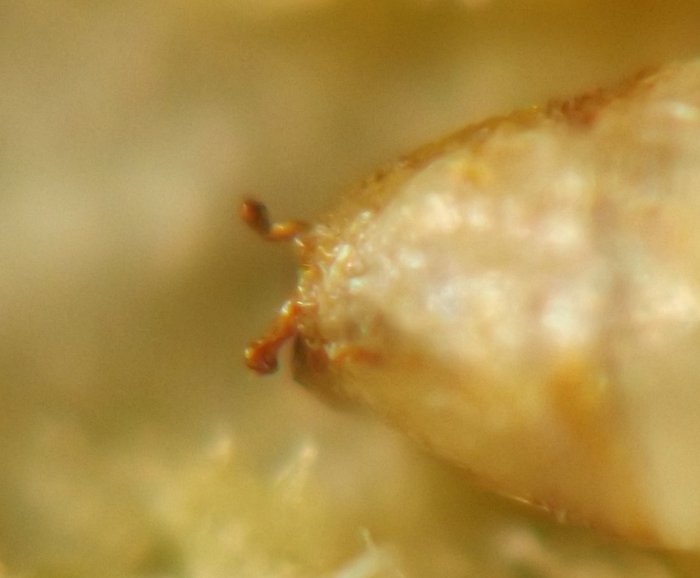
Posterior spiracles of spent puparium under bark.
IMG#: 0359-20
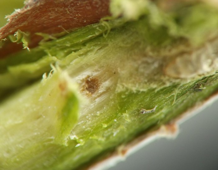
Puparium under bark at end of abbreviated shallow tunnel that originated deep in the stem interior; the point where the tunnel moved transversely from deep to shallow tissues has filled in with brownish callus tissue.
IMG#: 0359-21
Brownish callus tissue revealing origin point of tunnel from deep in stem interior.
IMG#: 0359-22
Region of stem immediately behind spent puparium, further dissected, showing connection between shallow pupation tunnel and larva's main area of tunneling activity deep in the stem interior.
IMG#: 0359-23
Tunneling in stem interior.
IMG#: 0359-15
Another example of a spent puparium under bark of the host.
IMG#: 0359-32
A third example of a spent puparium under bark of the host.
IMG#: 0359-33
Spent puparium under bark.
IMG#: 0359-35
Spent puparium under bark; as with the example in images 0359-16 through 0359-23, note the abbreviated shallow tunnel that originated from deep in the stem interior.
IMG#: 0359-36
Abbreviated shallow tunnel with spent puparium, and brownish callus tissue filling in the point where the tunnel emerges from deeper tissues.
IMG#: 0359-37
Region of stem immediately behind spent puparium, further dissected, showing connection between shallow pupation tunnel and larva's main area of tunneling activity deep in the stem interior.
IMG#: 0359-38
Region of stem immediately behind spent puparium, dissected, showing connection between shallow pupation tunnel and larva's main area of tunneling activity deep in the stem interior.
IMG#: 0359-39
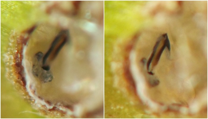
Two views of the cephalopharyngeal skeleton of a larva, left behind on the inner wall of the anterior end of the spent puparium.
IMG#: 0359-34
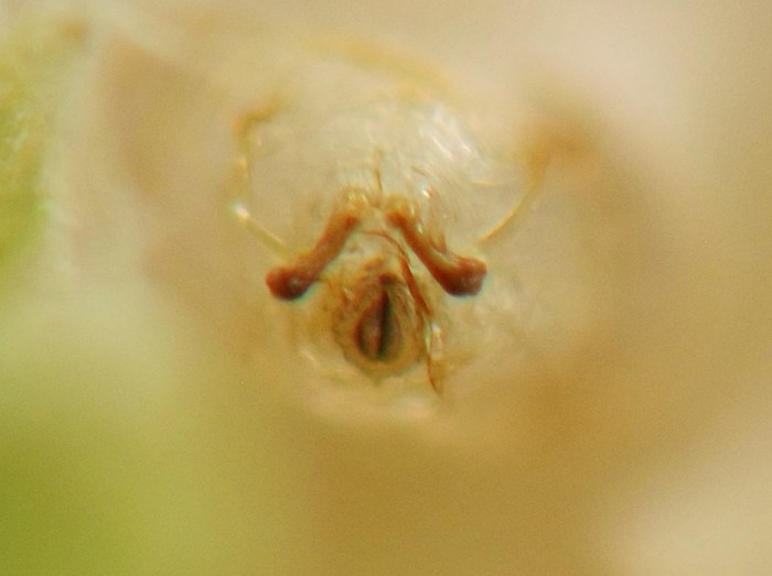
Rear view, posterior end of spent puparium.
IMG#: 0359-24
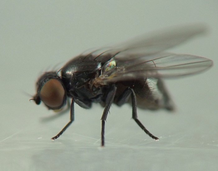
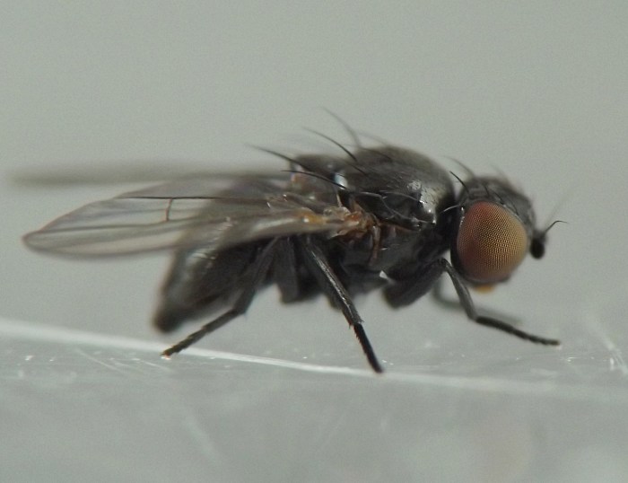
Reared adult, lateral. (Photo date: May 5, 2022)
IMG#: 0359-25
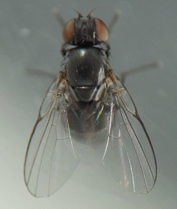
Reared adult, dorsal. (Photo date: May 5, 2023)
IMG#: 0359-26
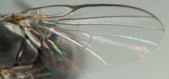
Wing of adult.
IMG#: 0359-27
Reared adult, lateral. (Photo date: May 4, 2022)

Specimen data for images

Coll. 08/19/18, photos same day (young larva, 01-04); coll. 02/20/18, photos same day (older larva stretched out straight in stem tunnel, 05-06); coll. 02/17/18, photos same day (older larva in stem, 07-12); coll. 04/09/22, photos same day (already-spent stem bump and a partial already-spent puparium under bark, 13-15); coll. 04/09/22 as intact puparium, adult em. 05/04/22, photos 05/04/22-05/05/22 (adult and puparium from which she emerged, 16-27); coll. 05/11/22, photo on 05/12/22 (already-spent stem bump, 28); coll. 05/11/22 as intact puparium, adult em. 05/21/22, photos 05/21/22 (puparium from which adult emerged in captivity, 29-39); coll. 02/01/18, photo taken on 02/02/18 (already-spent stem bump, 40).

References

Ellis, W.N. 2023. Phytobia cambii. In Plant Parasites of Europe [website]. Retrieved October 2, 2023 from here.

Page created 10/02/23. Last update: 11/17/24