Internal feeder (Lepidoptera: Heliozelidae) in Parthenocissus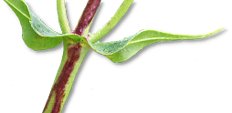
| Record no.: | 0363 |
|---|---|
| Feeding guild: | Internal feeder in petiole |
| Taxonomy: | Lepidoptera: Heliozelidae: cf. Heliozela sp. near aesella |
| Stages observed: | trace, larva |
| Distribution observed: | IA |
| Hosts in Parthenocissus: | undetermined P. sp. (woodbine / Virginia creeper) |
This unusual stem and petiole dweller was documented in the literature via a brief note by Weiss and West (1925), who initially collected it in New Jersey in 1921. They were able to rear larvae through to the pupal stage, but did not obtain adults. McGiffen and Neunzig (1985) conducted further documentation and rearing efforts. The following account expands on these authors' work, documenting several details of the immature life history with photographs and notes.
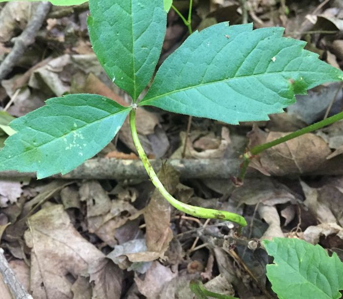
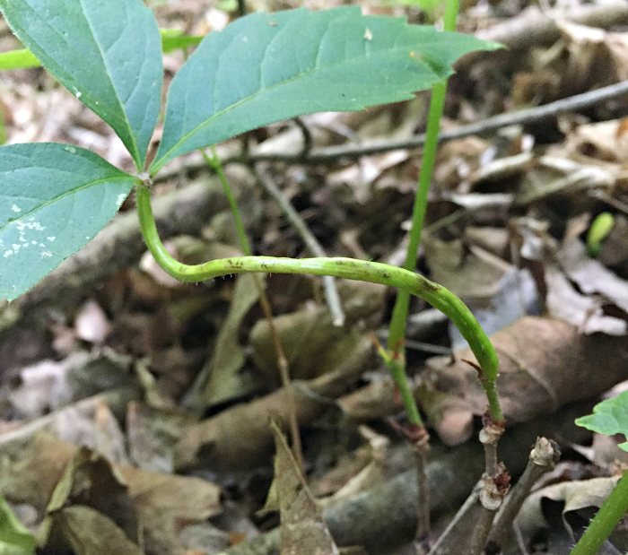
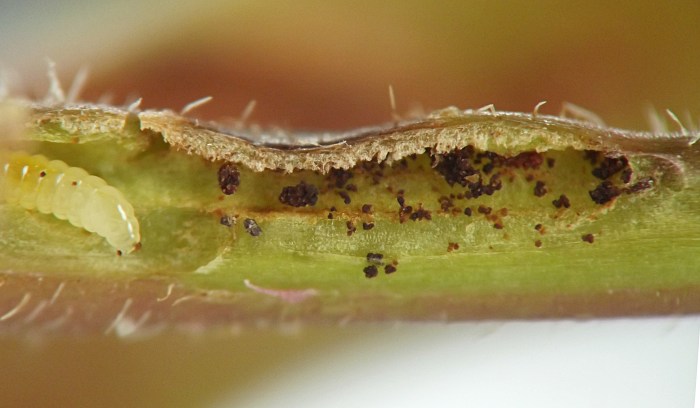
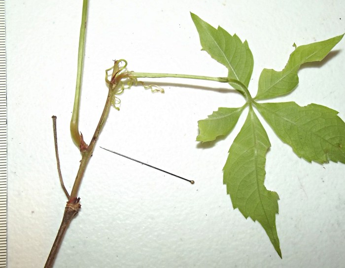
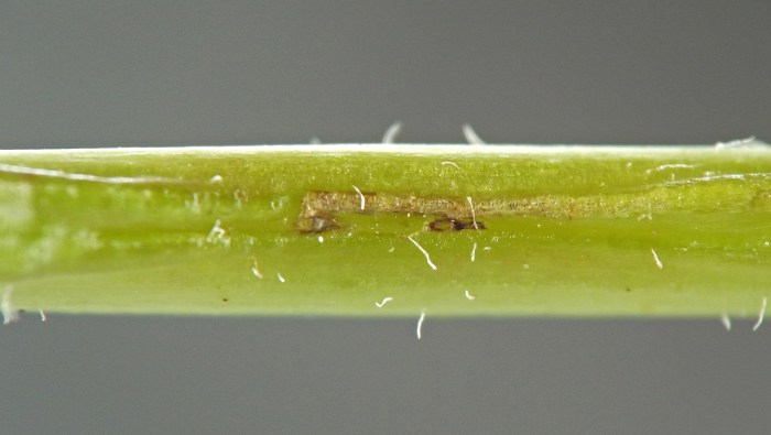
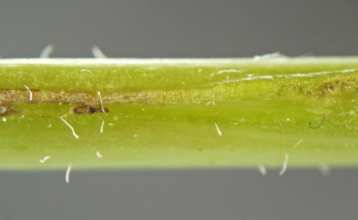
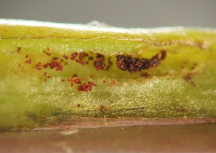
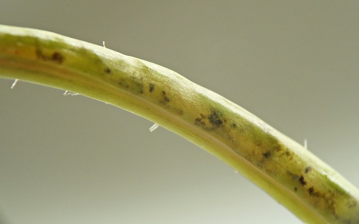
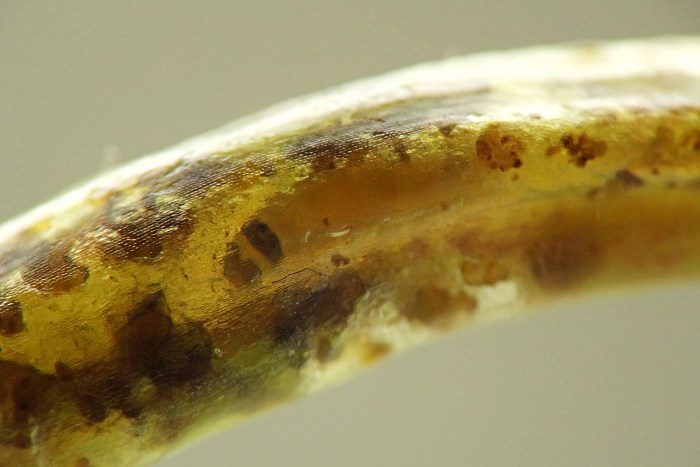
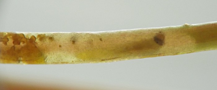
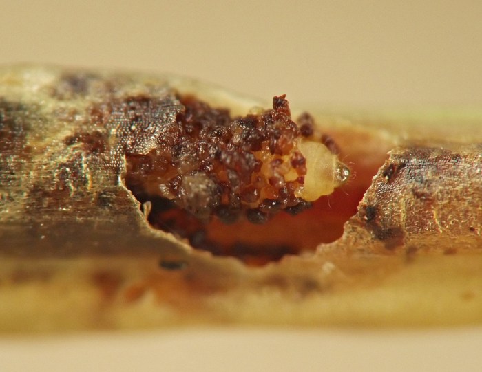
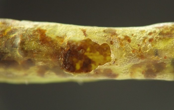
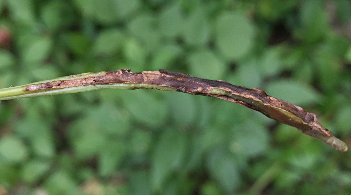
The first ready indicator of this internal feeder consists of subtle longitudinal swellings on petioles and new stems of the host, which appear in late May and early June. The galls stretch along the length of the petiole or stem and may extend partway around the circumference of these plant parts, but in most examples I saw in 2023, there remained at least one unaffected strip of tissue along the length of the plant part.
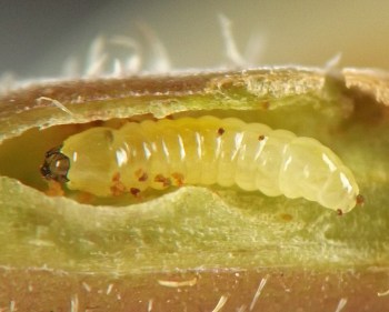
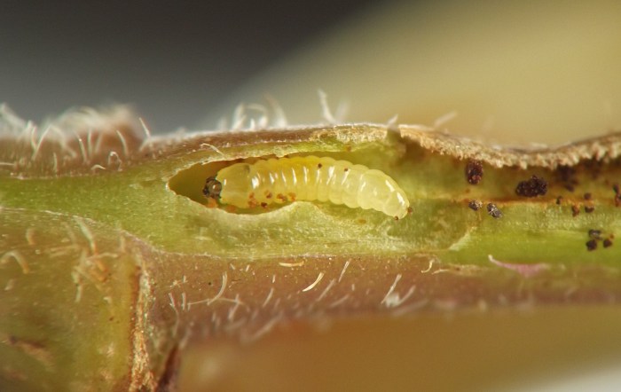
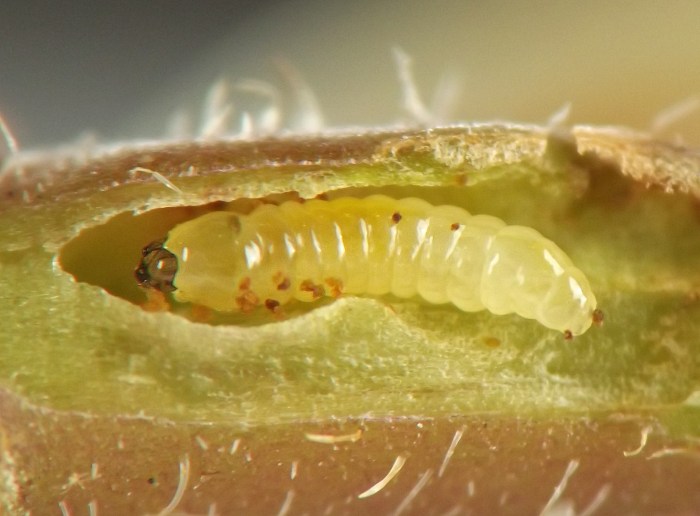
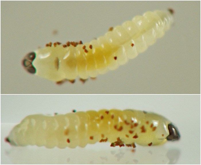
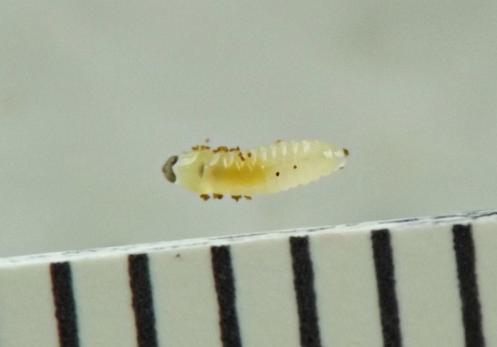
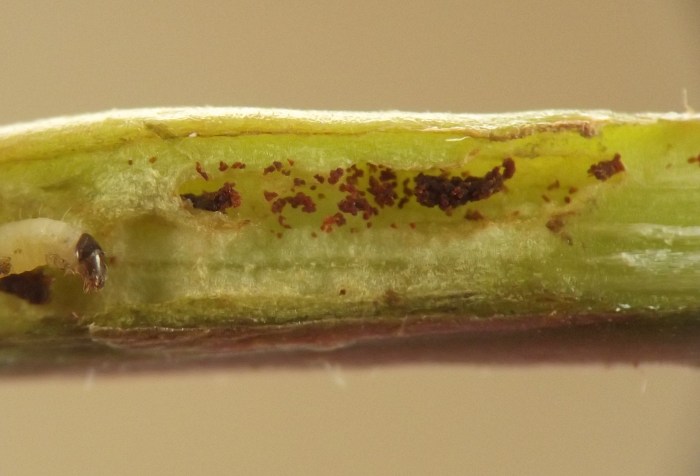
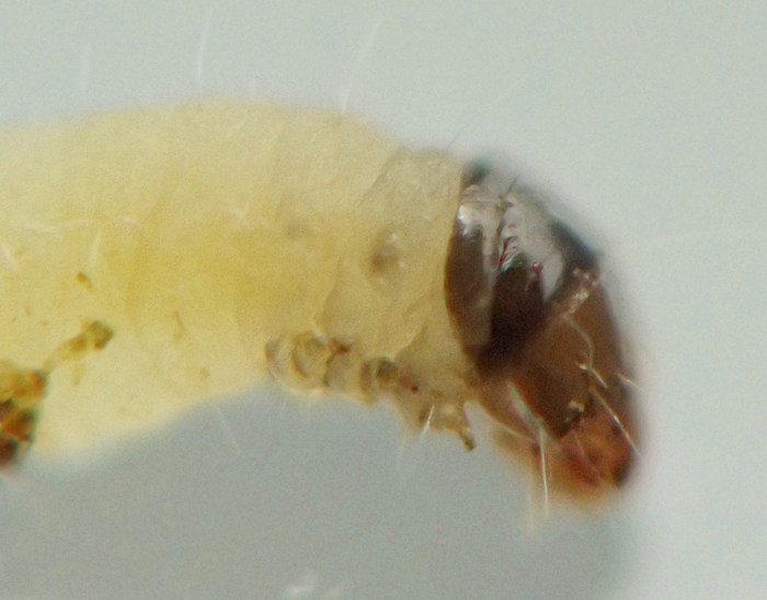
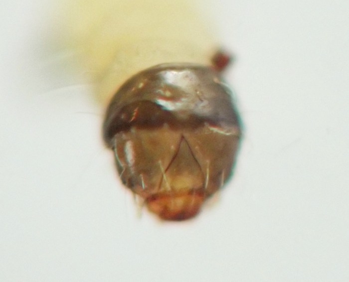
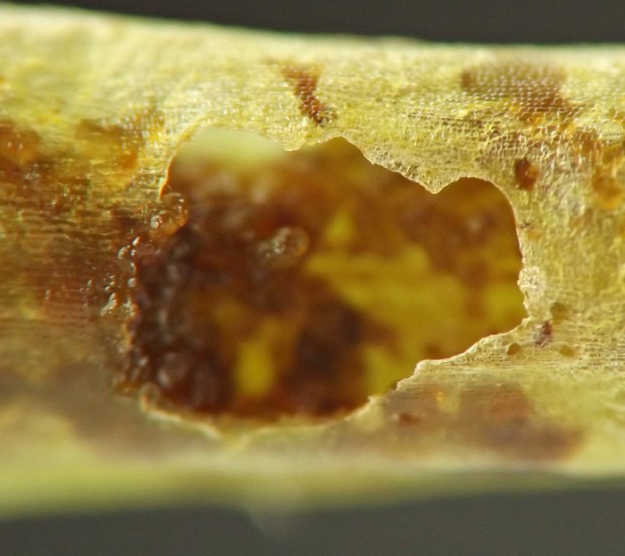
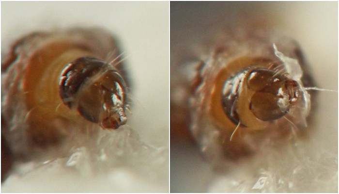
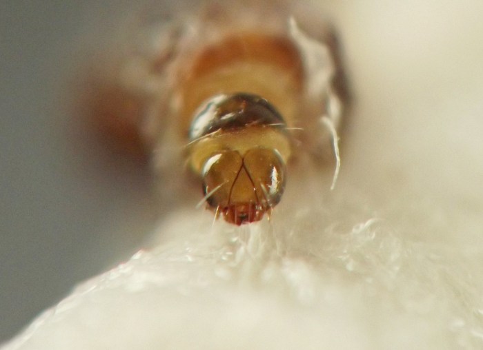
A gall dissected on 24 May 2022 contained no visible larvae -- perhaps because eggs laid in the tissue had not yet hatched. In 2023, starting on June 3, dissecting some of these galls revealed lepidopteran larvae tunneling inside them and producing reddish-brown solid frass in the tunnels. At that time two different forms of larvae could be found in the galls, a smaller form with a dark head capsule but without obvious thoracic legs, and a slightly larger form with a dark head capsule, a dark thoracic shield, and well-developed thoracic legs. McGiffen and Neunzig reported that "larvae of an unidentified weevil" were mixed in with the Heliozela larvae in the petiole, peduncle, and/or shoot galls they examined, and stated that "[the] weevil larvae can be distinguished from Heliozela larvae by the weevils' lack of thoracic legs and crochets" (p. 52). I did not observe any larvae that clearly belonged to Coleoptera in the galls I examined, and my strong impression was that the smaller, apparently legless larvae and the larger larvae with thoracic legs from these galls represented different instars of the same species, with the apparently legless form being an earlier instar, suggesting some form of advanced metamorphosis occurs from the legless to the legged instar.
Multiple larvae could be found in the galls affecting a single petiole or section of stem. One petiole I photographed contained at least three larvae.
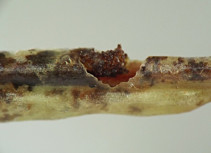
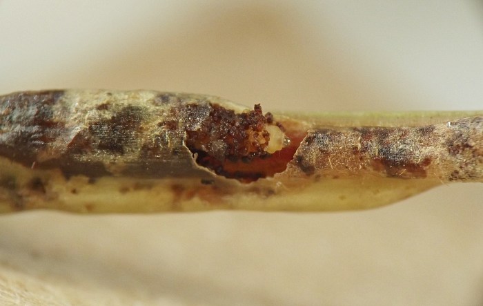
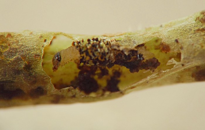
Larvae kept in containers indoors developed quickly, excavating extensive tunnel systems in the galls. Eventually only the second, larger larval form could be found in the plant material. The tunneling soon destroyed most of the tissue in the swollen portions of the petioles and stems, causing the galls to darken due to the internal accumulation of dark brown frass visible through the plant epidermis. Larvae could also be observed through the plant epidermis as they adjusted position, fed, and defecated. I watched larvae interact with one another inside the tunnels and engage in housekeeping by moving particles of frass around with their mandibles.
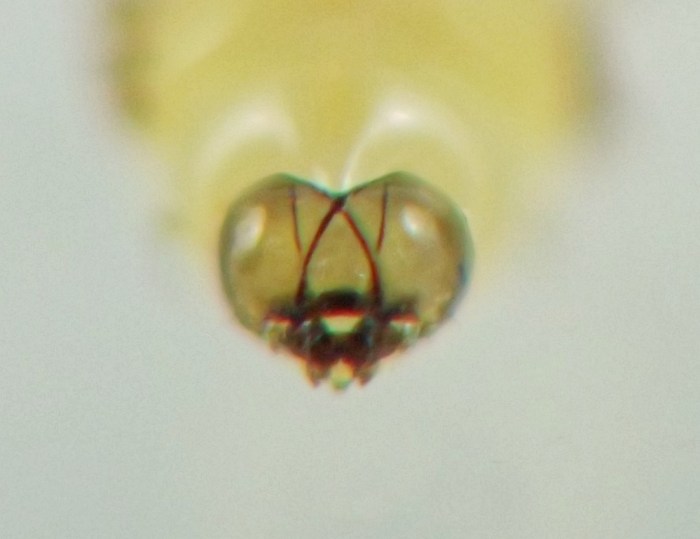
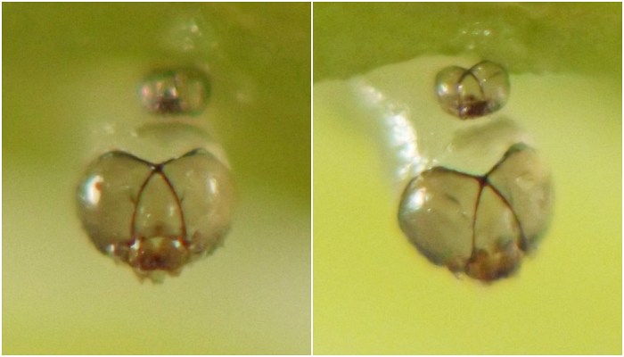
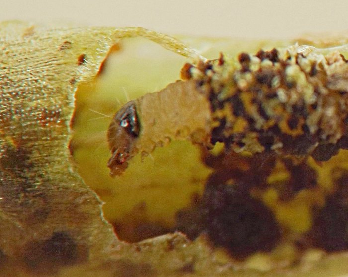
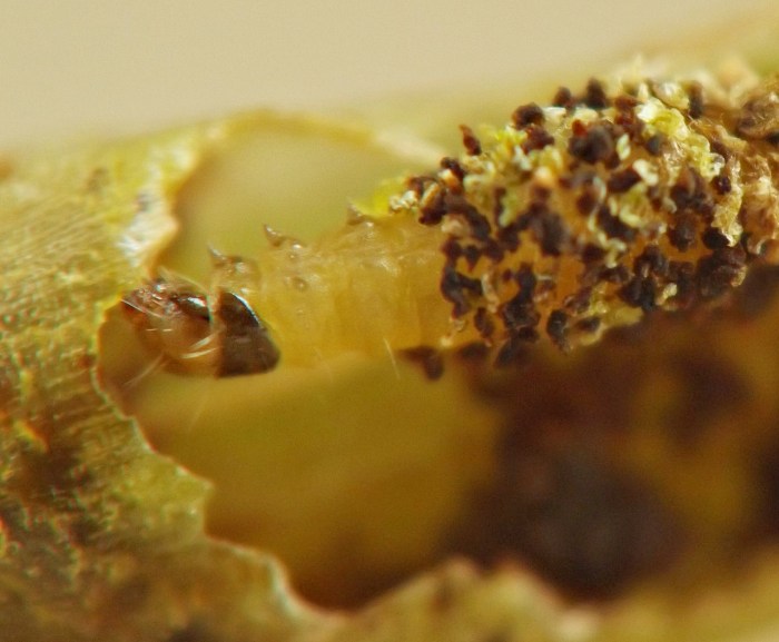
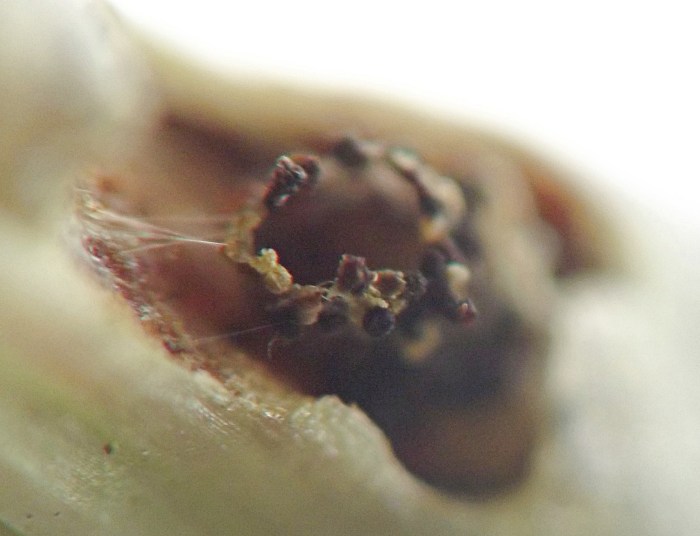
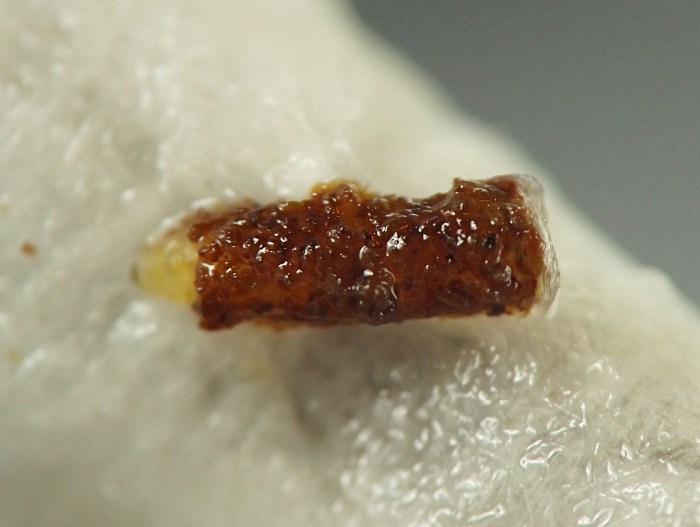
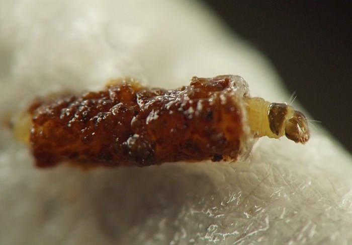
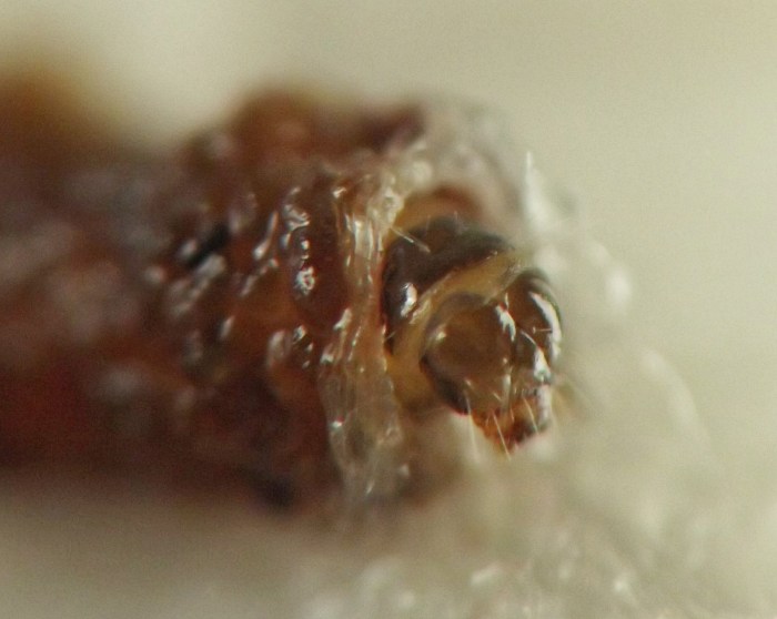
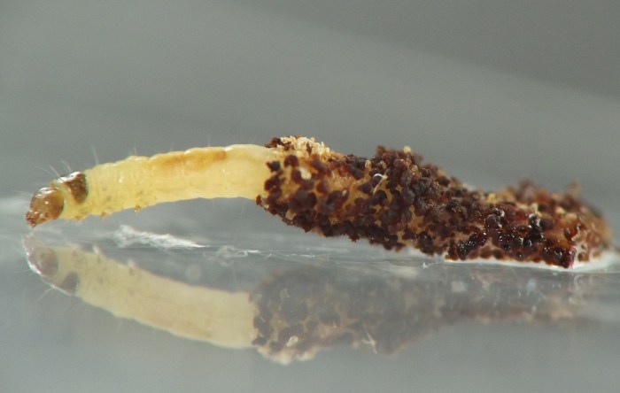
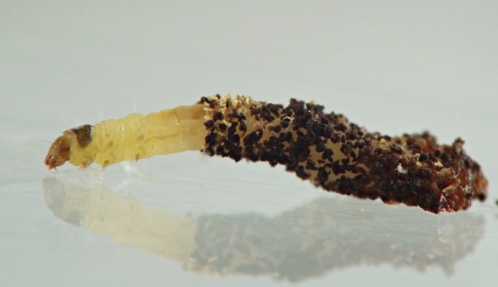
Eventually, while still inside the galls, larvae begin building cases for themselves. From June 6-8, 2023, I found several larvae dwelling within nearly complete cases beneath gaping holes they had created in the plant epidermis. These larvae attached to their cases not only bits of frass but also small excised pieces of stem or petiole epidermis. They cut out the fragments of epidermis with their mandibles, and manipulated the pieces of frass with their mandibles as well. These building materials were attached to the case with silk.
Prior authors have indicated or implied that this interwoven composite of frass and silk comprises the base layer of the case, and that the large holes in the plant epidermis occur due to the eventual disintegration of the injured plant tissue and are not directly related to case construction. For example, Weiss and West wrote that their larvae constructed for themselves "case(s) made from particles of excrement" and then exited the galls "through small openings which later become ragged tears due to the collapse of the tissue" (p. 117). Similarly, McGiffen and Neunzig reported that the larvae they observed built "tubes of frass" around themselves and that a "plug of gall tissue" was not present in the pupal case, unlike for H. aesella larvae from leaf galls with whom such a plug is present; in addition, "after larvae have exited, the thin layers of the gall crack open" (p. 52).
However, my photos of larval cases suggest that the base layer of the case may actually consist mainly of plant epidermis that the larva cuts out and assembles into a tube, before it begins to add the numerous small fragments of frass and epidermis to the exterior of the case. This base layer is especially noticeable when the air is dry and the small fragments on the case exterior have desiccated, revealing the layer underneath (see photos). The excision of such a large quantity of epidermis as the foundation of the case could help explain the large size of the hole in the epidermis that accompanies some larvae and their cases. Still, I cannot absolutely confirm this hypothesis because I did not directly observe the beginning of case construction. If the case foundation is indeed constructed of epidermis, it's unclear to me whether it consists mainly of a single large piece or several smaller pieces woven together.
According to Weiss and West, the larvae "crawl beneath the debris on the forest floor" after leaving the plant and then pupate "within the case during the last of August." (They did not describe how they were able to observe the forest floor behavior or how they cared for their captive larvae over the course of the summer until the larvae pupated.) Despite multiple attempts, Weiss and West were unable to succesfully obtain adults from overwintered pupae. They shared larval specimens with Carl Heinrich, who "identified the larva as that of a species of Adelidae, possibly Adela ridingsella." I do not know which characters Heinrich used to arrive at the family-level identification of Adelidae or what if anything led the authors to believe the species might be A. ridingsella. McGiffen and Neunzig refer to this moth as Heliozela sp. near aesella, and I rely on this later determination of theirs herein.
As far as I know, as of 2023 no adults have yet been reared.
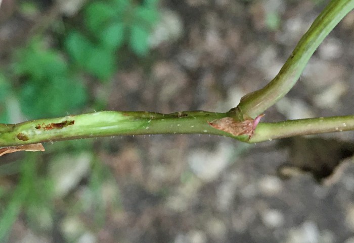
In mid-June, an apparent Heliozela species tunnels in somewhat older main stems of Parthenocissus in my study area (record 0729). Aside from the subtle swelling shown by some inhabited nodes, this borer's presence is generally not externally obvious until the larva finishes feeding and establishes a gaping oval hole in the outer wall of the stem, through which it evacuates the plant material, also while ensconced in a case partly composed of frass. In 2025, I found a recently hatched larva of this insect on 11 June, and it formed a case around itself and exited the plant material in the rearing container by 19 June, with the oval exit holes of other individuals formed in the field by 20 June. Because of the differences in phenology and plant damage, I believe this may be a different species than the one described in the current report, and I have given it a different record number.
 Prev
Prev
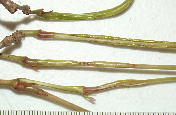
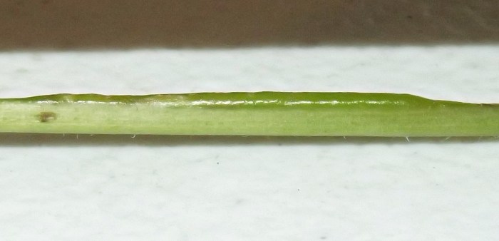
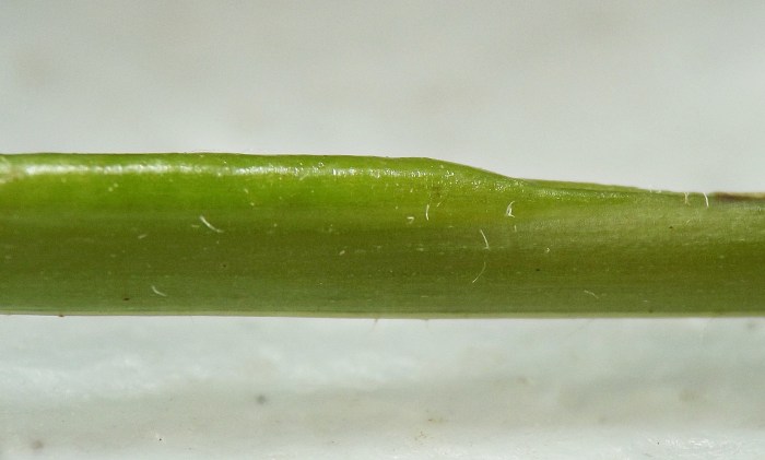
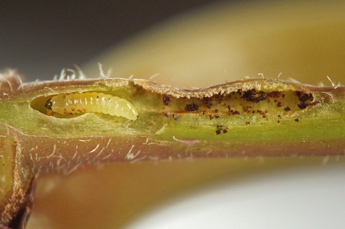
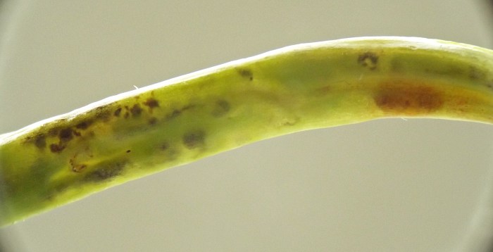
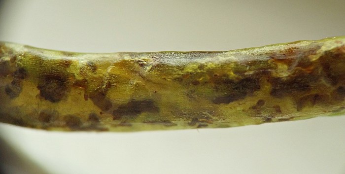
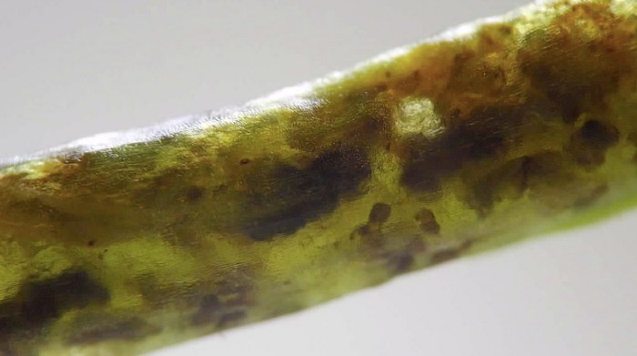
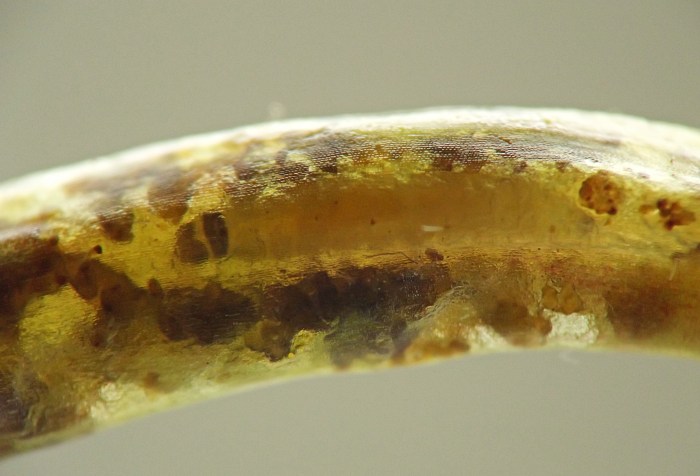
Field photos taken 06/05/23 (01-02); field photo taken 06/03/23 (03); coll. 06/05/23, photo taken 06/06/23 (04); coll. 06/03/23, photos taken 06/03/23-06/04/23 (05-21); coll. 06/05/23, larvae forming cases and exiting galls starting 06/07/23, photos taken 06/06/23 through 06/08/23 (22-44); field photo taken 08/08/23 (45).
- McGiffen, K.C. and H.H. Neunzig. 1985. A guide to the identification and biology of insects feeding on muscadine and bunch grapes in North Carolina. North Carolina Agricultural Research Service, Bulletin 470.[return to in-text citation]
- Weiss, H.B. and E. West. 1925. An adelid gall on Virginia creeper (Lepidoptera). Entomological News 36(4):116-118. Retrieved September 7, 2023 from https://www.biodiversitylibrary.org/page/24738162.[return to in-text citation]
Page created: September 6, 2023. Last update: June 9, 2026