Tunnel walls are distinctly ragged as with other dipteran borers
Pupation is external, off the plant, with the puparium very small (~2mm in length) and yellow, reminiscent of Liriomyza
The population of this borer I observed in 2023 suffered very high larval mortality, with roughly 66-75% of examined larvae having been killed by a parasitoid wasp or otherwise dying of natural causes before reaching the puparium stage
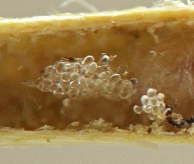
In a few cases I observed, after the agromyzid larva had been consumed by a parasitoid wasp larva, the most visible remnant of the agromyzid larva was a cluster of calcareous discs left over from the interior of the larva -- a body part the parasitoid was evidently unable or unwilling to eat. For more about such discs, see Ellis (2023)
Some of the parasitoid wasp larvae spun cocoons in the tunnels not far from their hosts' remains
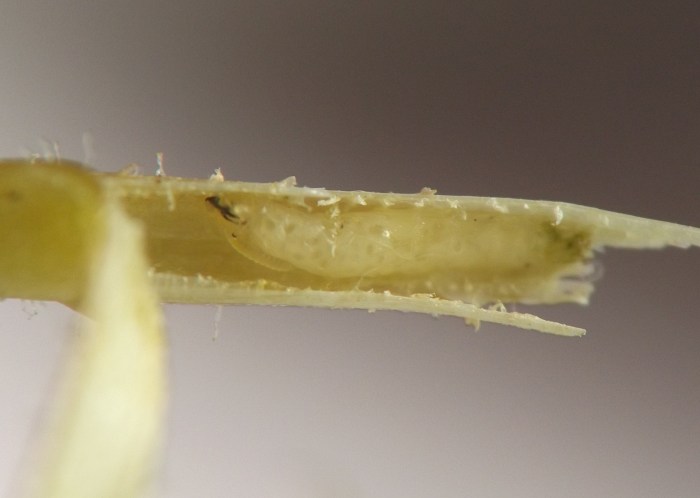
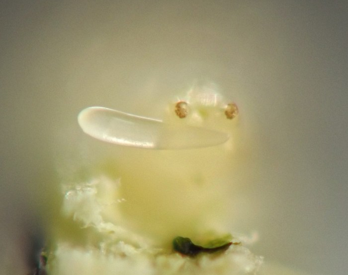
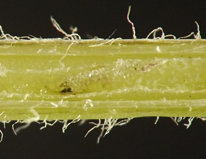
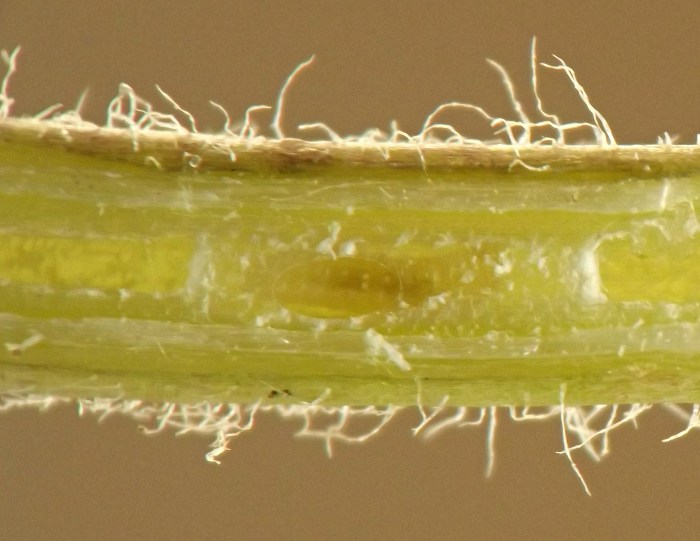
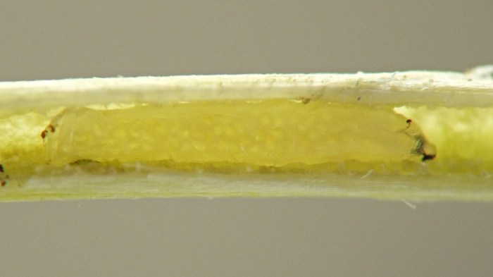
Image #: 376-02Parasitized borer larva in stem. The parasitoid is visible just below the borer larva's cephalopharyngeal skeleton, clinging to its integument. (Photo date: June 21, 2023)
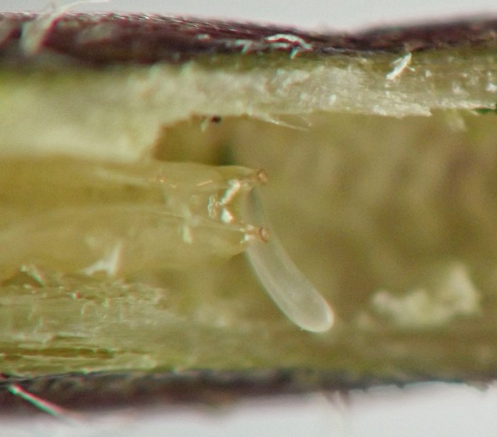
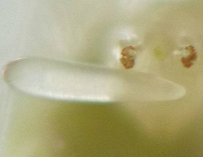
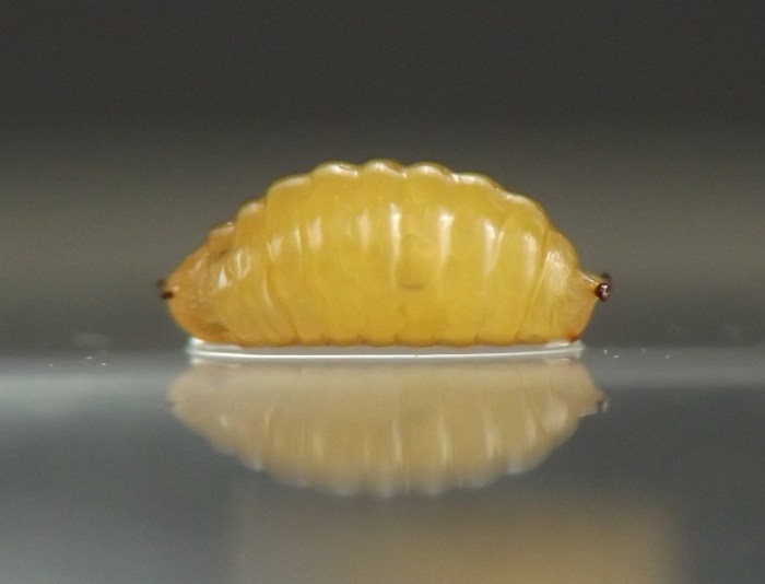
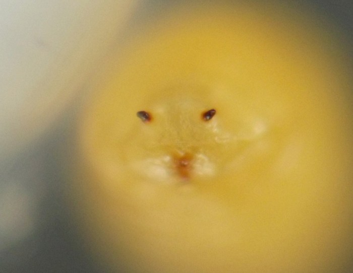
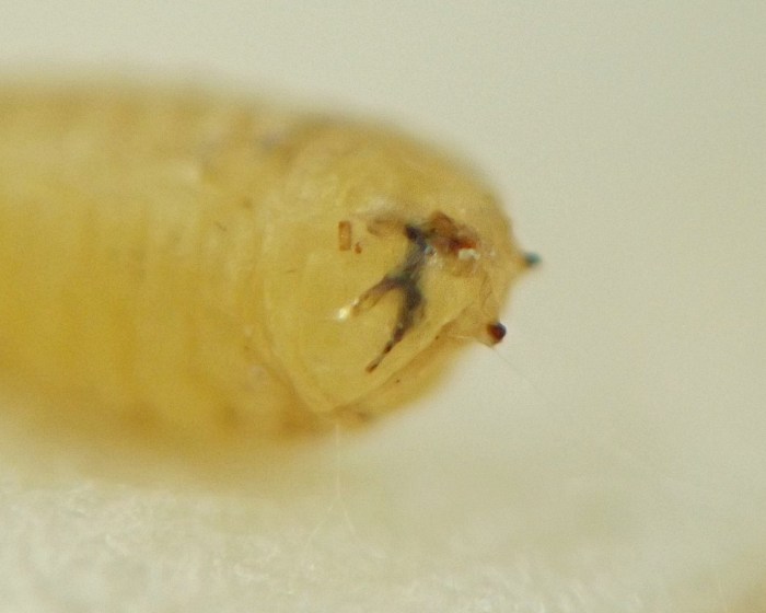
Image #: 376-07Detail, rear view of larva's posterior end, with apparent parasitoid life stage attached. Note also the characteristics of the posterior spiracle that is more or less in focus to the right of center.
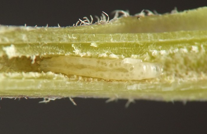
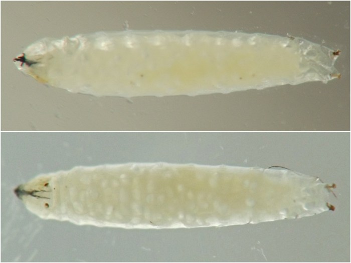
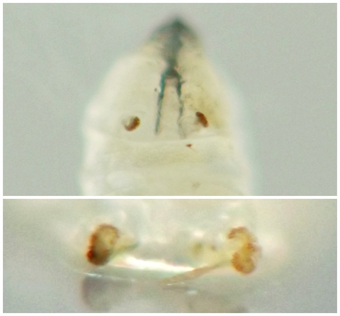
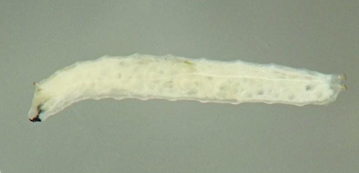
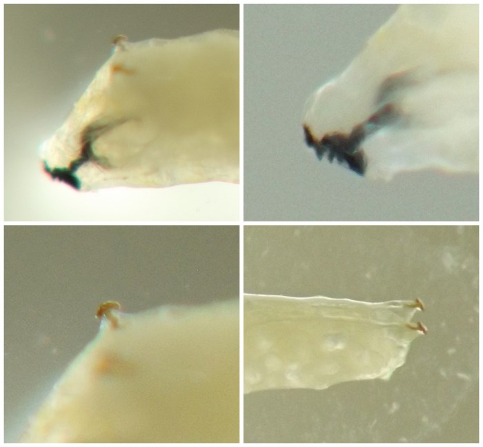
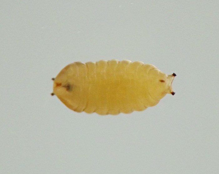
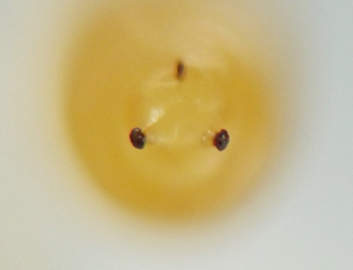
Image #: 376-11Several views of a larva. Top left: larva's anterior end, showing cephalopharyngeal skeleton; top right: detail, front of cephalopharyngeal skeleton, showing teeth on mouthhooks; bottom left: anterior spiracle; bottom right: rear half of larva, showing posterior spiracles in dorsolateral view.
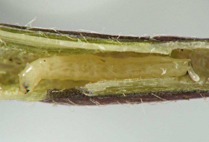
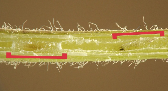
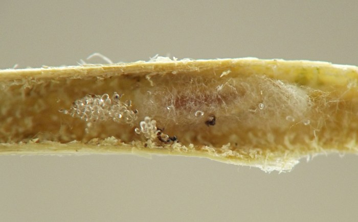
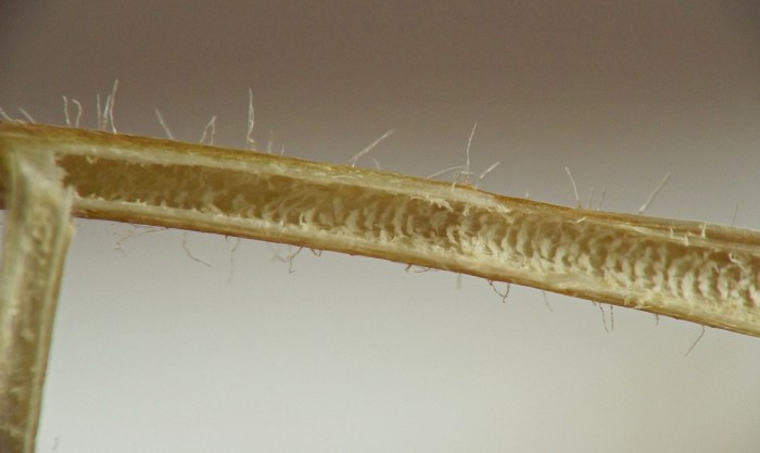
Image #: 376-12An affected stem containing remains of an agromyzid borer larva (right bracket) with the parasitoid larva that consumed the agromyzid nearby (left bracket), in a cocoon it has spun in its host's tunnel.
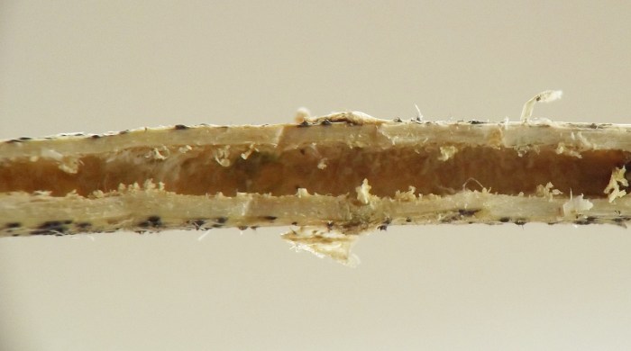
Image #: 376-15A second example of a tunneled stem containing the remains of the borer larva with its parasitoid in a cocoon nearby. Here, the most visible remnant of the agromyzid larva is the cluster of calcareous discs left over from the interior of the larva -- a body part the parasitoid was evidently unable or unwilling to eat. For more about such discs, see Ellis (2023). Just to the right of the discs in this image is the parasitoid larva, faintly visible through the thin wall of its spun cocoon.
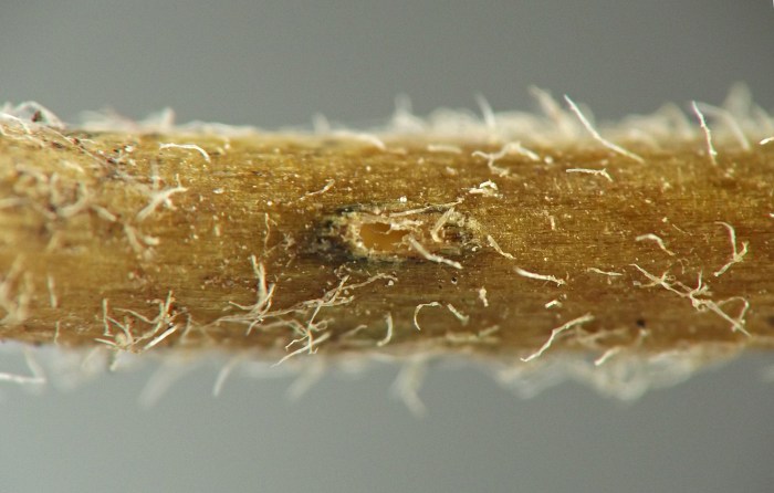
Image #: 376-17Larvae of this borer exit the stem to pupate. This image shows the exit hole left by a fully mature larva when it vacated the stem in order to pupate in the soil.
Image #: 376-25Puparium, after overwintering, with the cephaloskeleton of the final-instar larva appressed to the inner wall of the puparium on the anterior end.
Coll. 06/29/23, photos taken 06/29/23-06/30/23 (01, 03, 12-17, 19); coll. 06/21/23, photos taken 06/21/23-06/22/23 (02, 04-11, 18); coll. 06/29/23, puparium found outside of stem in rearing container on 07/01/23, photos on 07/02/23 (20-24); coll. 06/29/23, puparium by 07/xx/23, puparium photographed after overwintering on 03/17/24 (25-26).
 Prev
Prev