Thrips (Thysanoptera) associated with cavities in plant tissue
by J. van der Linden
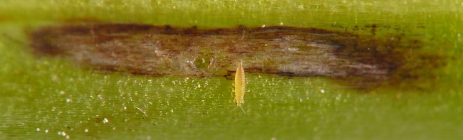
24 July 2026
In: Van der Linden, J. 2026. Upper Midwest Stem Insect Survey. Self-published Web reference.
Available online at https://insect-pages.github.io/survey/pages/thrips.html.
Summary of findings
Between 2022 and 2026, I observed thrips inside short, linear, subepidermal cavities in stems of bluebells (Mertensia virginica); inside natural hollows in midribs and petioles of jewelweed (Impatiens sp.); and under the separated epidermis of a petiole of Virginia waterleaf (Hydrophyllum virginianum).
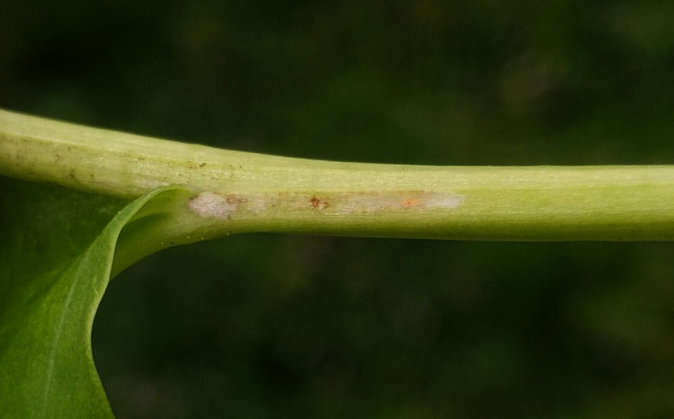
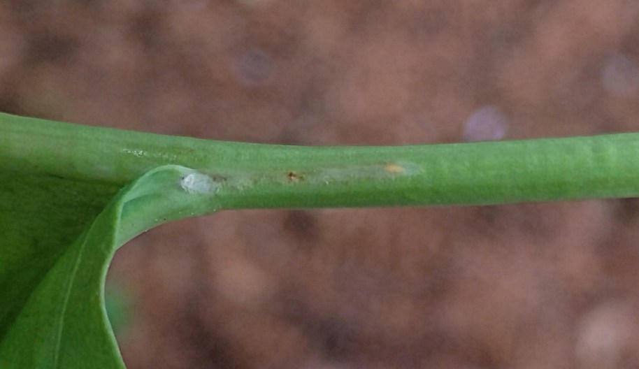
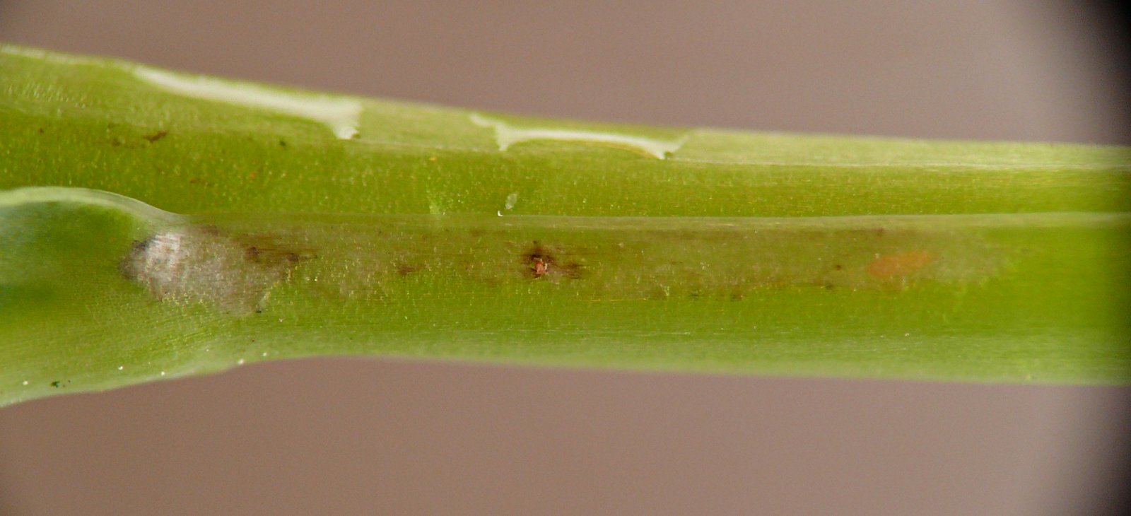
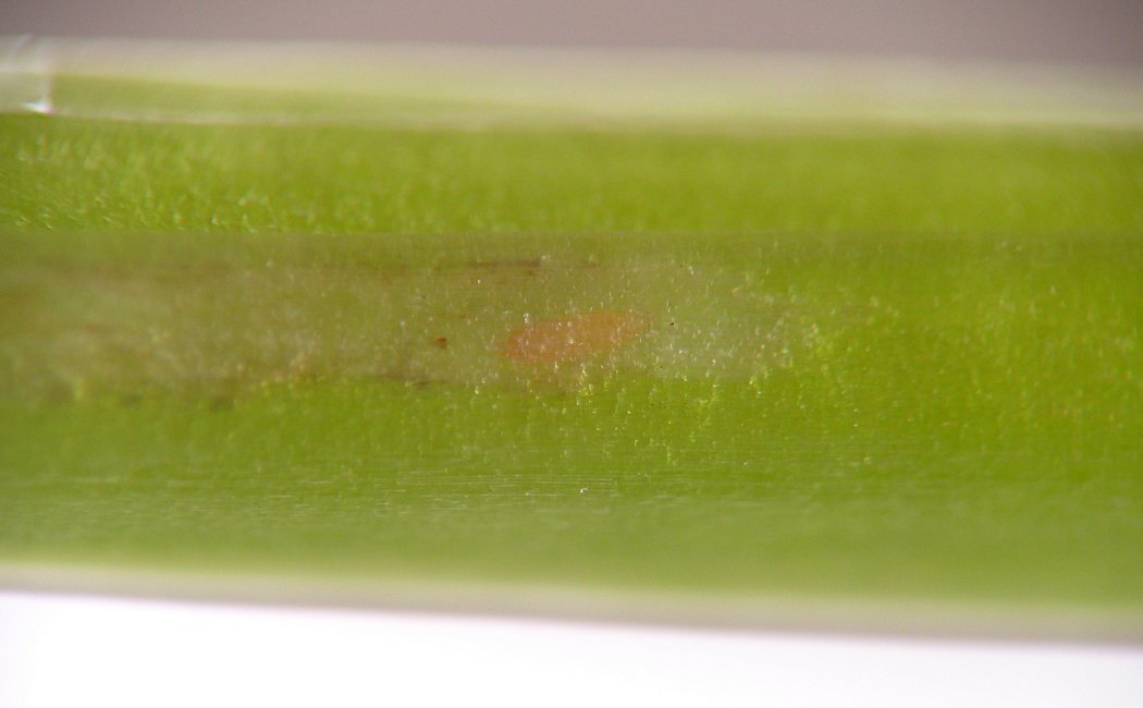
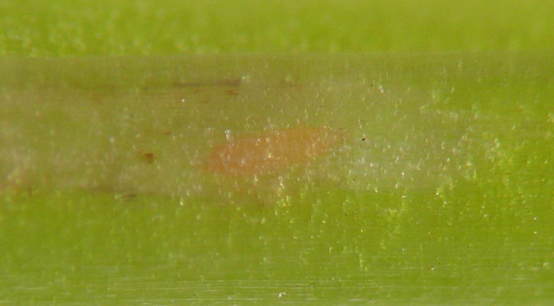
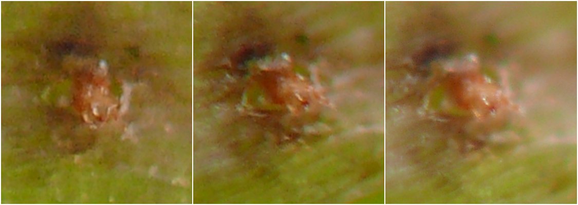
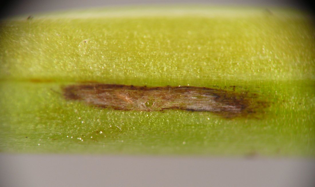
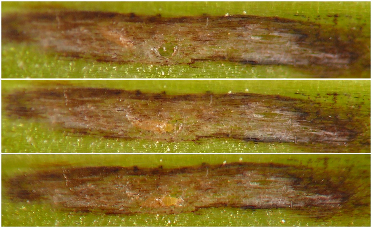
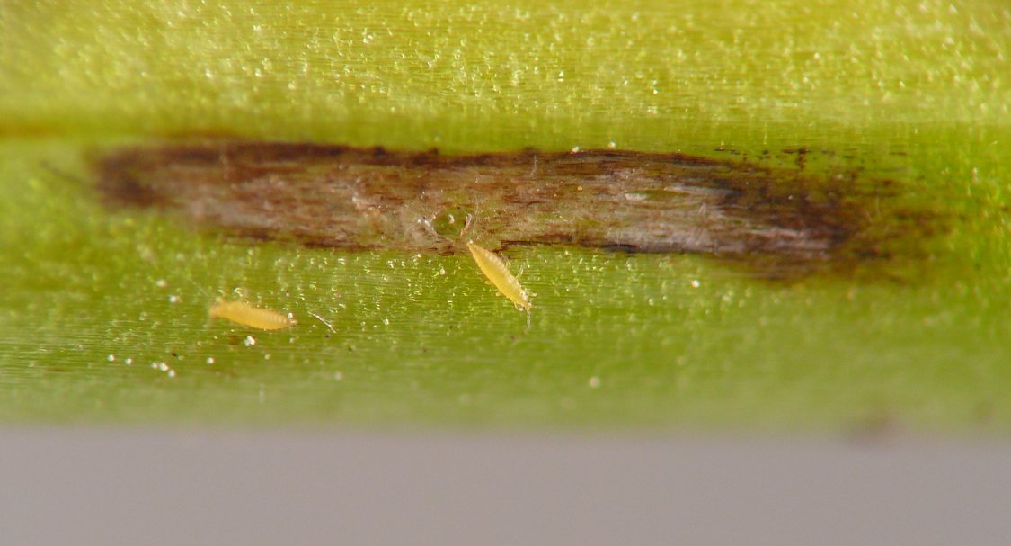
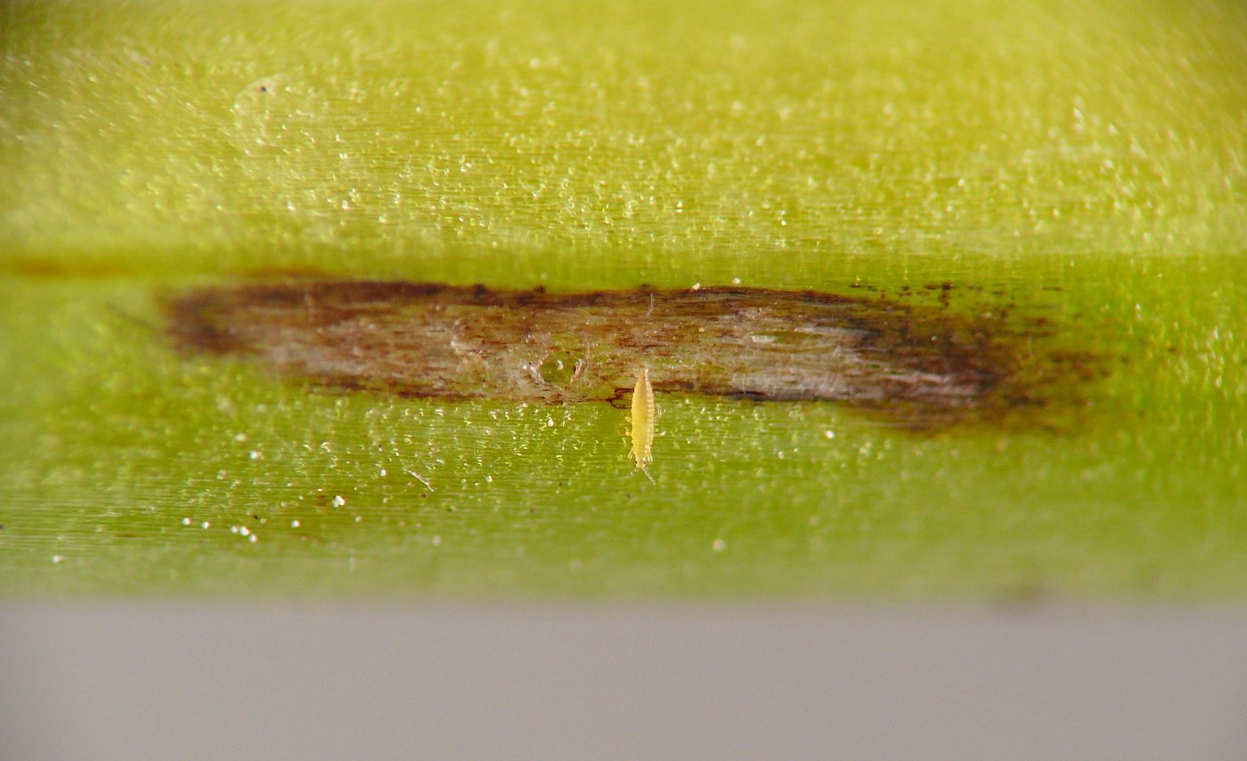
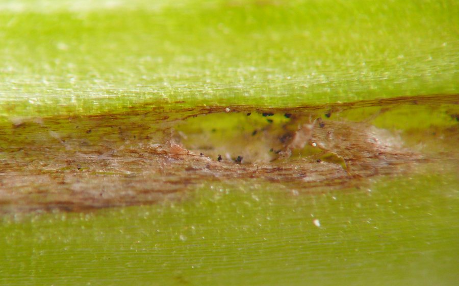
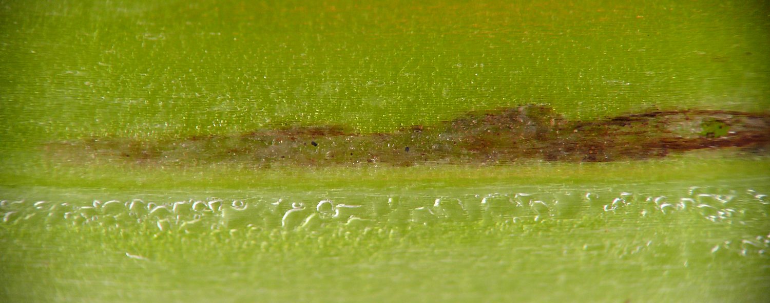
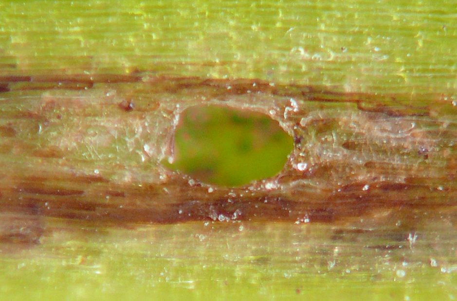
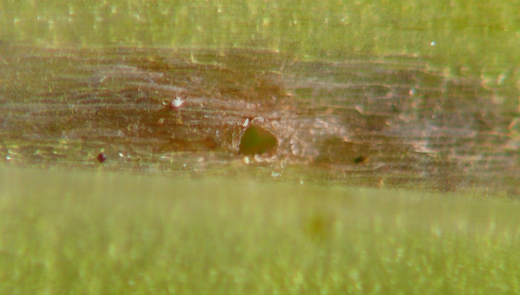
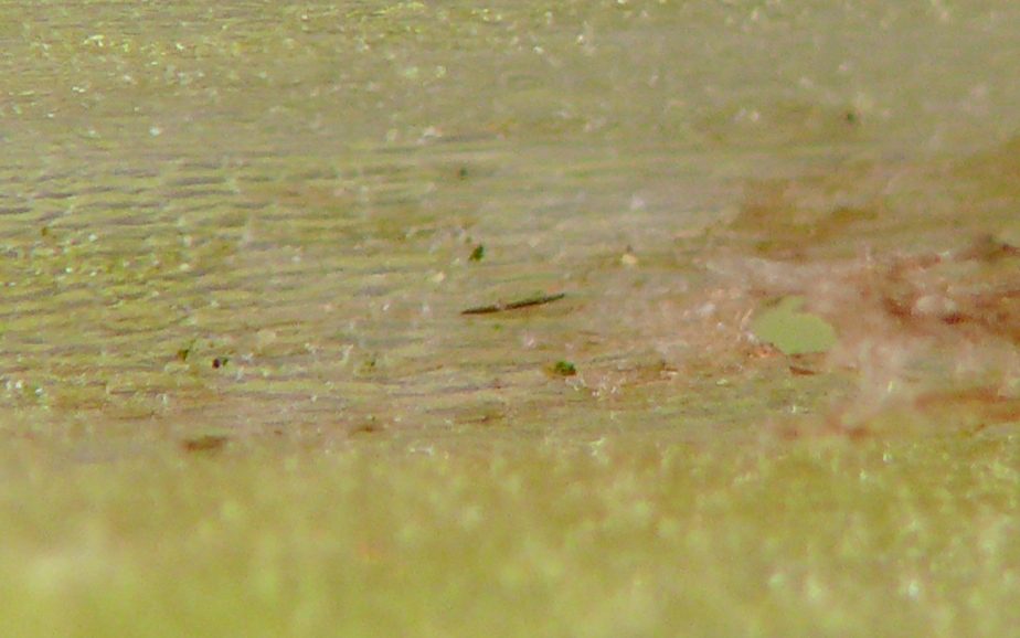
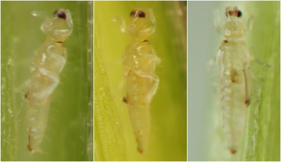
Typically some type of perforation existed in the epidermis of the stem or stemlike structure through which the thrips could theoretically enter or exit the cavity or expel debris from it, and in a few instances I saw evidence of them using such holes for one or more of these purposes. However, the full context for the thrips' association with these epidermal holes and the connected, inhabited cavities in the plant tissue was not always clear. In one case on Mertensia virginica (images 01-07 & 56, below), a single thrips was positioned at the end of a linear cavity that appeared very fresh, with its head facing the apparent leading edge of the cavity, no other insects present, and the only entrance or exit blocked by (tentatively identified) thrips exuviae, suggesting this individual may have been the initial colonizer of this region of plant tissue, although I was unable to conclusively confirm this. On jewelweed, the leaf midribs and petioles are naturally hollow, and I have seen one case of a very young tortricid caterpillar (probably Pristerognatha agilana) living inside a petiole and a few other cases where an apparent caterpillar tunnel led from the petiole into the main stem, indicating that thrips are not the only insect that may take up residence inside the midribs and petioles. However, in several jewelweed midribs and petioles actively inhabited by thrips and not by caterpillars, careful examination revealed no tunnel leading from the petiole into the main stem and no other obvious place through which an insect the size of a caterpillar could have exited the leaf. This strongly suggested to me that at least some endophytic thrips in the jewelweed midribs and petioles are not simply inquilines in former caterpillar tunnels.
Affected cavities on all three plants usually contained some degree of blackening of the inner walls, which was sometimes externally visible through the plant epidermis. I interpreted this blackening as sign of feeding by the thrips; however, in most cases I was not able to discern clear evidence of frass accumulation. Hering (1951) reported an example of thrips ejecting frass through epidermal holes in an agromyzid mine, suggesting that a relative dearth of obvious frass in a plant cavity inhabited by thrips is probably not solid evidence that no feeding has taken place, especially if one or more epidermal holes are present.
In Mertensia virginica
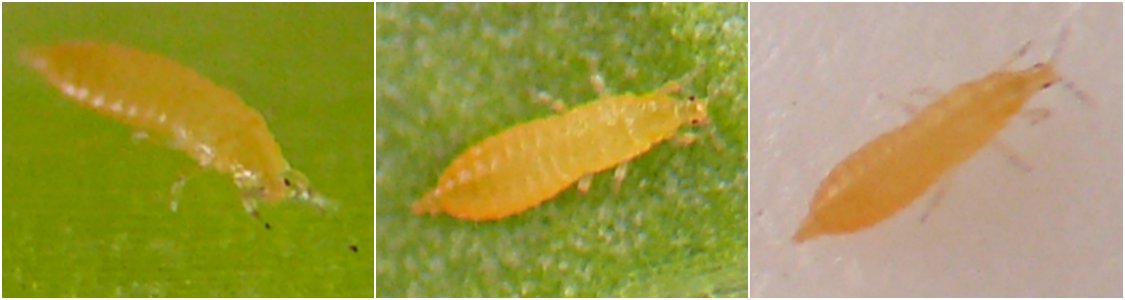
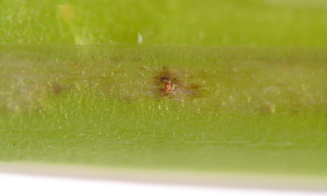
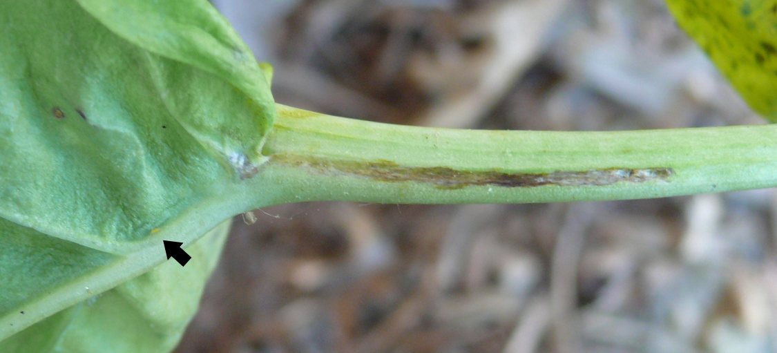
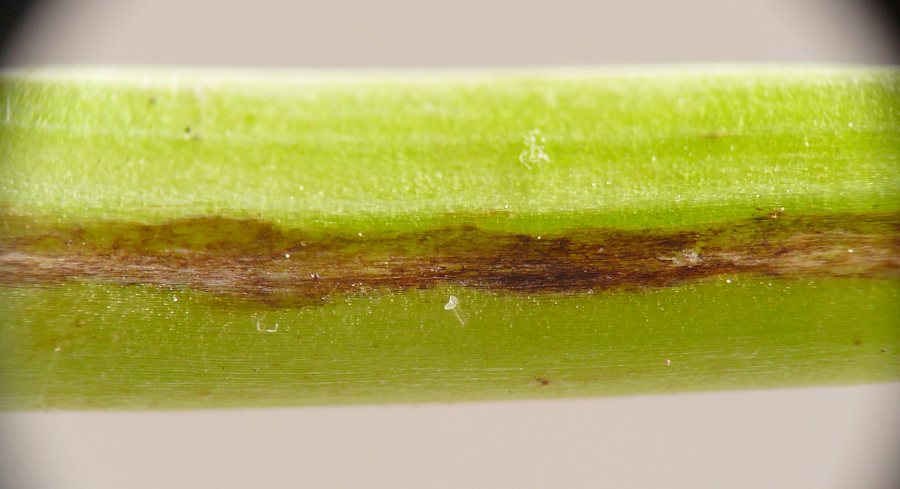
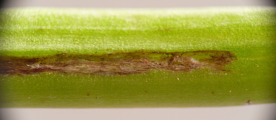
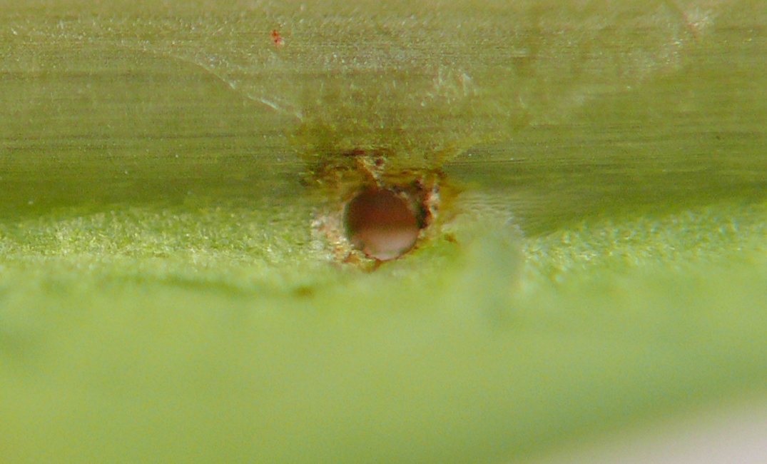
I found two examples of short, linear, subepidermal cavities actively inhabited by thrips, and several additional cavities with similar structural characteristics but no thrips present. In most cases, the single epidermal hole was positioned approximately halfway between the two ends of the cavity, although I did find an instance where the hole was located at one end. In one of the actively inhabited examples (images 01-07 & 56, below), a small reddish object plugged the central hole. Photos show paired, elongate-conical, banded microstructures on the object, suggesting it could represent the exuviae of the thrips in the cavity, whose legs were shaped and colored similarly to the microstructures, but I was unable to confirm this hypothesis.


In Impatiens
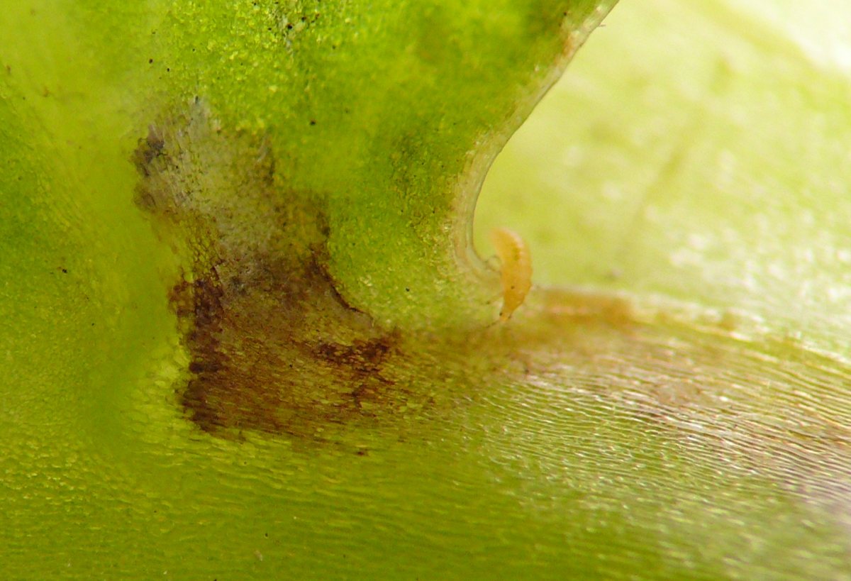
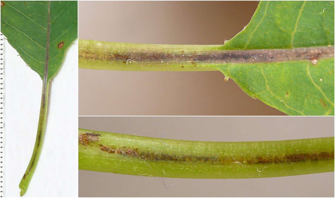
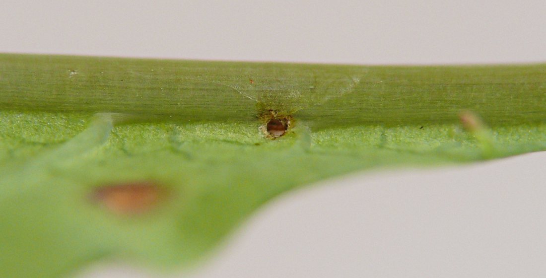
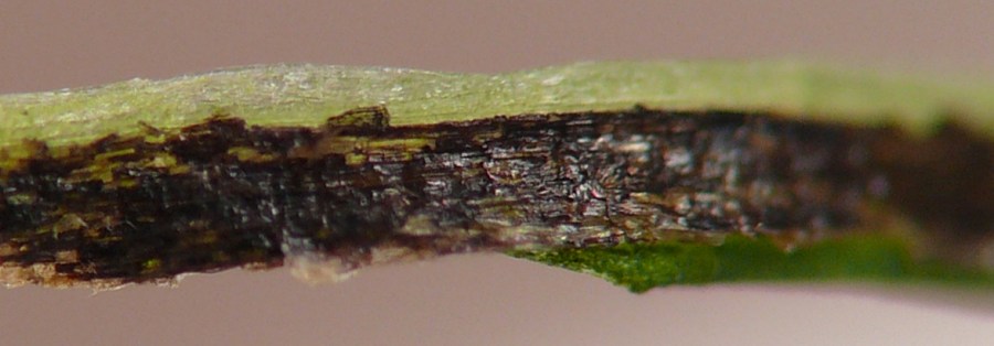
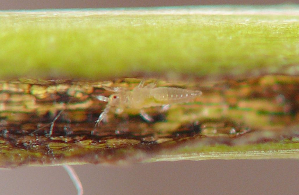
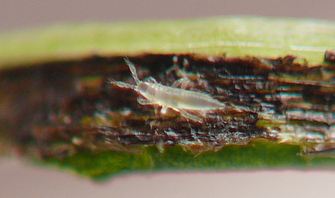
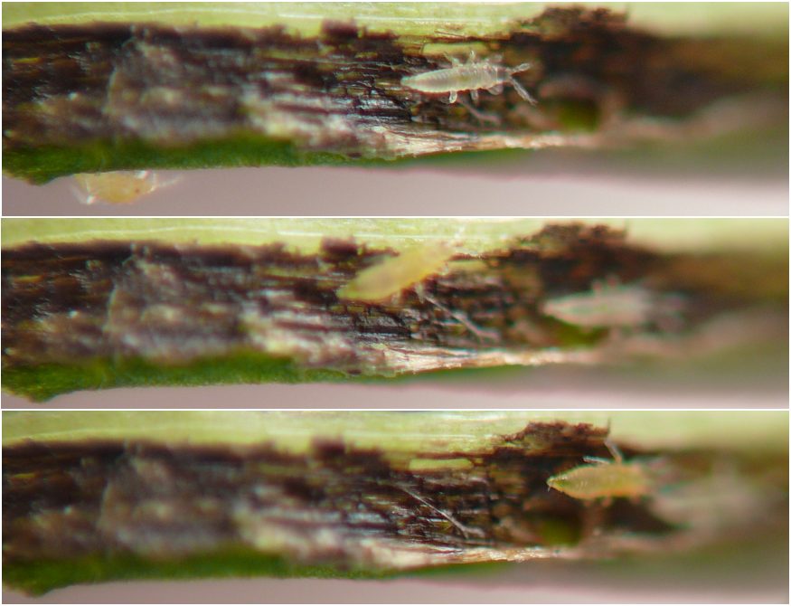
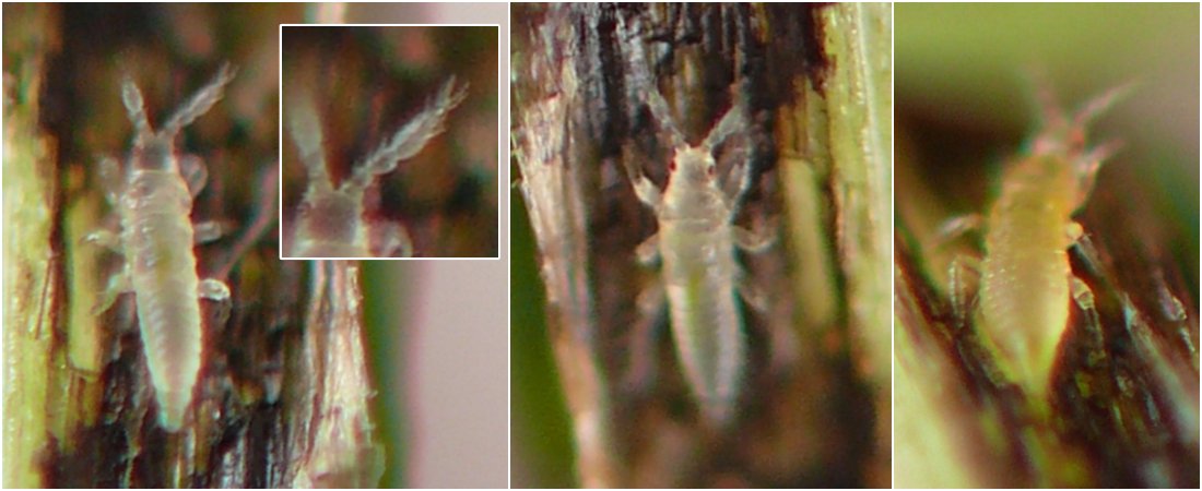
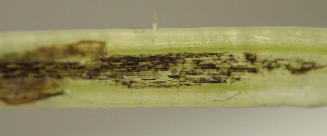
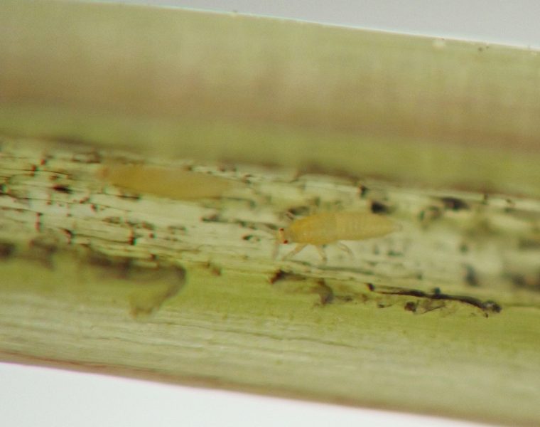
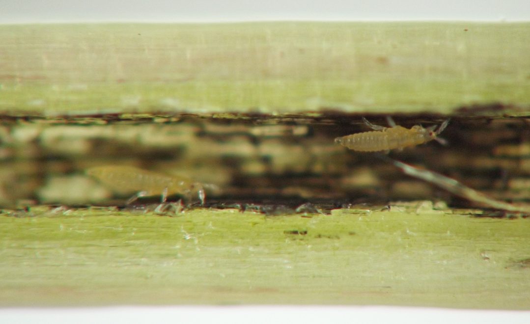
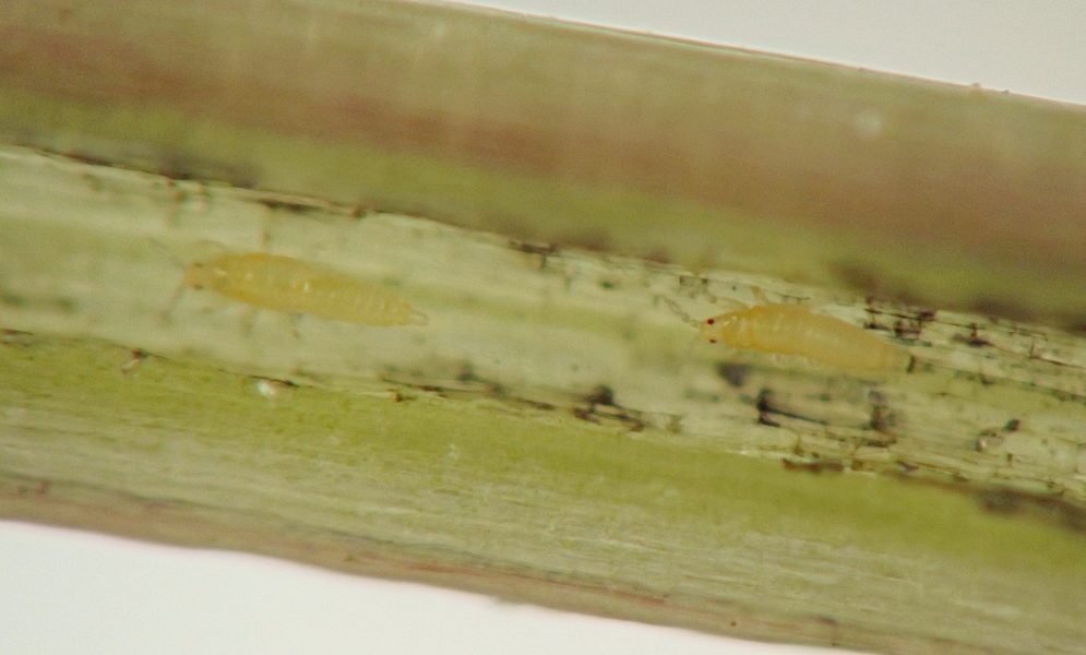
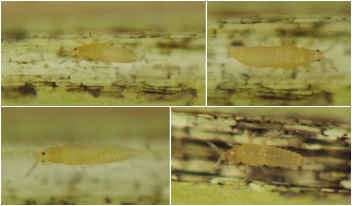
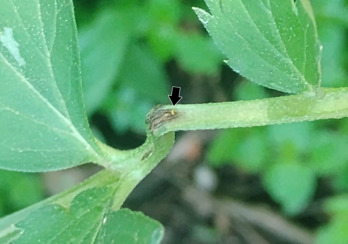
I have found numerous examples of jewelweed midribs and petioles inhabited by thrips. The leaf stalks are naturally hollow, and those with thrips inside usually show some externally visible darkening of the inner walls of the hollow, which I have attributed tentatively to the feeding action of the thrips, although solid frass is usually not visible.
The following excerpts from my journal provide some additional details.
- Late June, 2023: "[I] was able to find just one affected [jewelweed] leaf that had some blackish discoloration to the midrib/petiole...It had a couple of small holes in the upper midrib/petiole epidermis that looked as though they were the result of something making dozens of small chews and eventually chewing their way in. There were only thrips in this petiole. That was the point where I wondered, could thrips be the culprit here, and only thrips, nothing else? But I didn’t think thrips had chewing mouthparts."
- Mid-July, 2023: "Thrips in inwardly discolored midrib/petiole, yellow jewelweed, Impatiens [pallida]...There was one extremely tiny, raised hole in the ventral petiole but no other disturbance to the outer layers of midrib/petiole tissue and no other way something could have entered or exited. Thus it seems likely the thrips was the original cause of the inner discoloration."
- Mid-July, 2023: "Tortricid larva inside midrib and petiole of leaf, jewelweed, Impatiens sp....There was some externally visible internal blackening of the petiole and midrib as a result of the larva’s presence. This is the first evidence I’ve seen that such blackening may be caused by the tortricid borer."
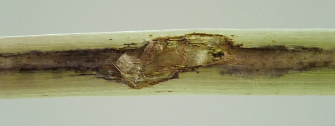
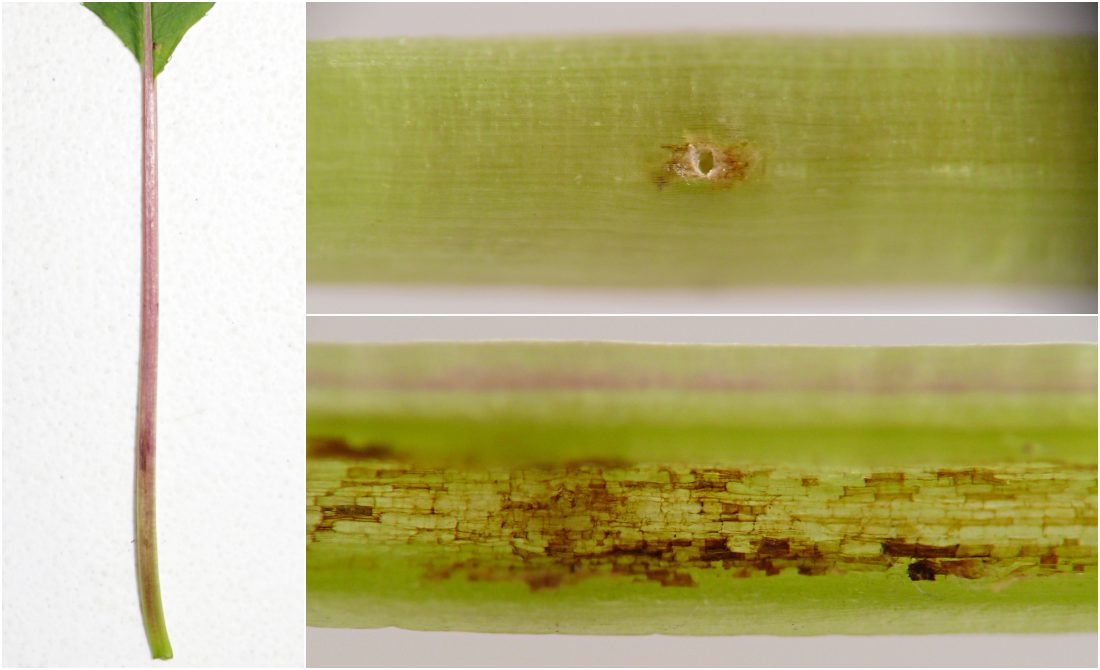
For comparison, I have also included images of midrib and petiole damage I believe was exclusively caused by the tortricid (image 55). In this case, what appeared to be an oviposition hole penetrated the underside of the petiole, and perhaps more tellingly, a brownish tunnel led from the naturally hollow petiole interior through the normally solid petiole base and into the main stem of the plant. These features were not present in several of the midribs and petioles I observed that were inhabited by thrips but not by caterpillars, suggesting to me that these thrips were primary colonizers of the plant tissue rather than inquilines of the tortricid.
In Hydrophyllum virginianum
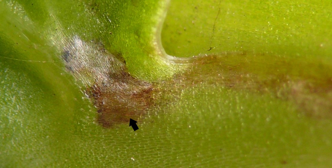
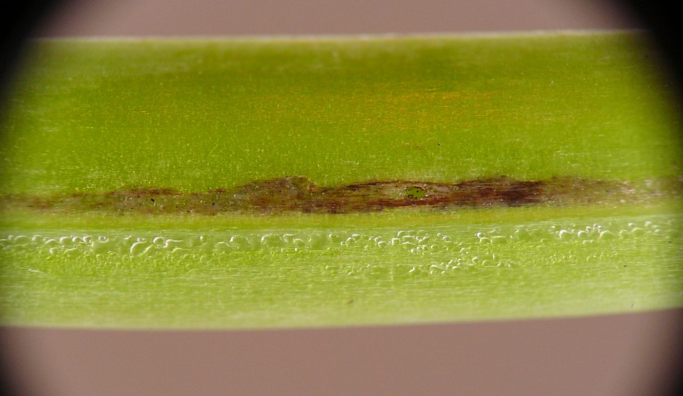
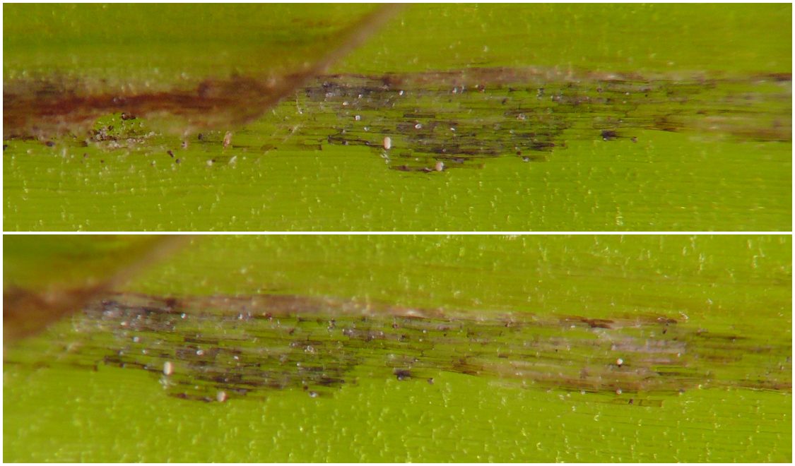
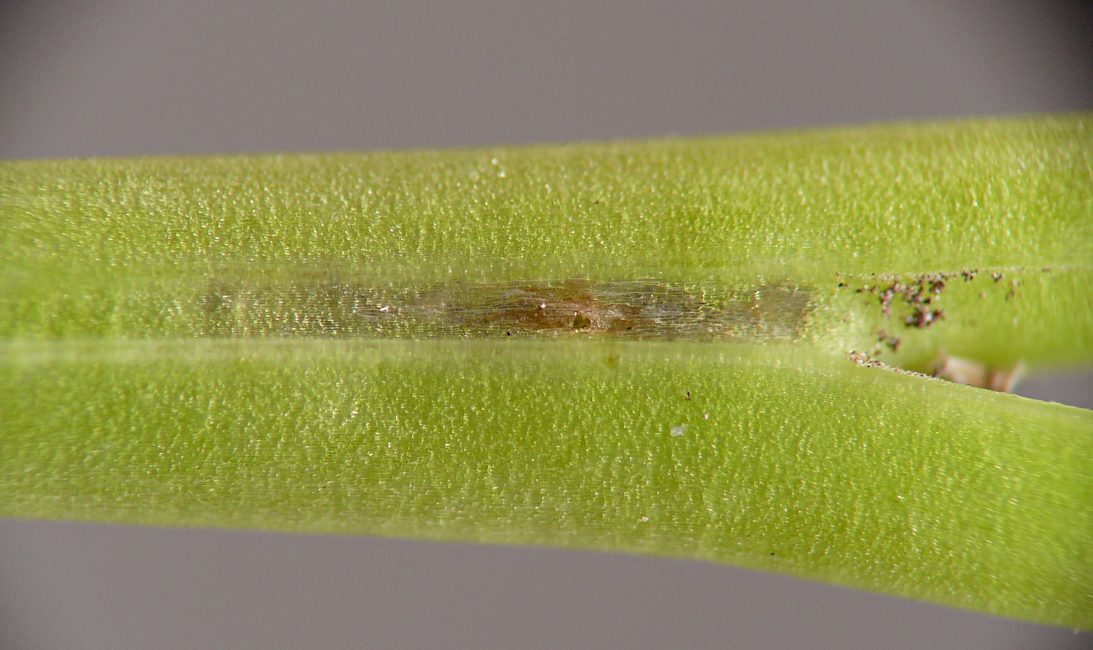
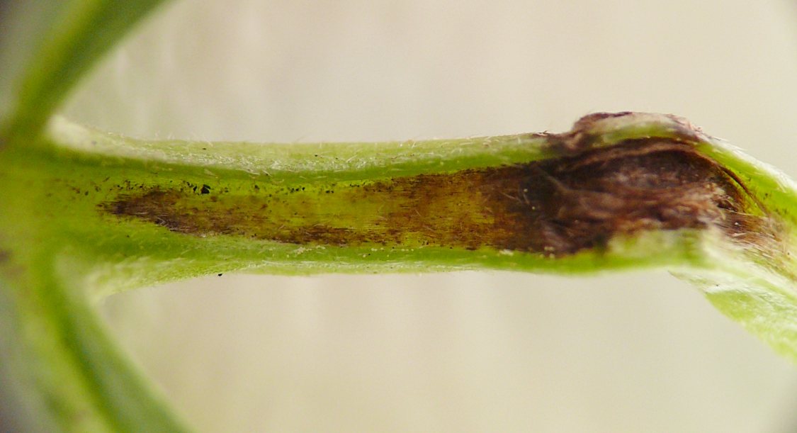
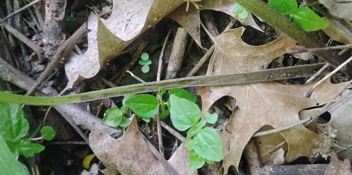
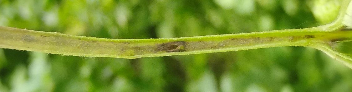
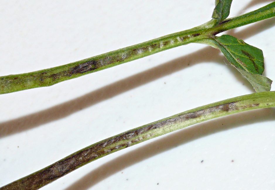
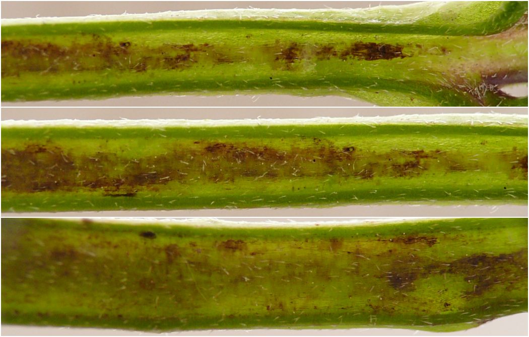
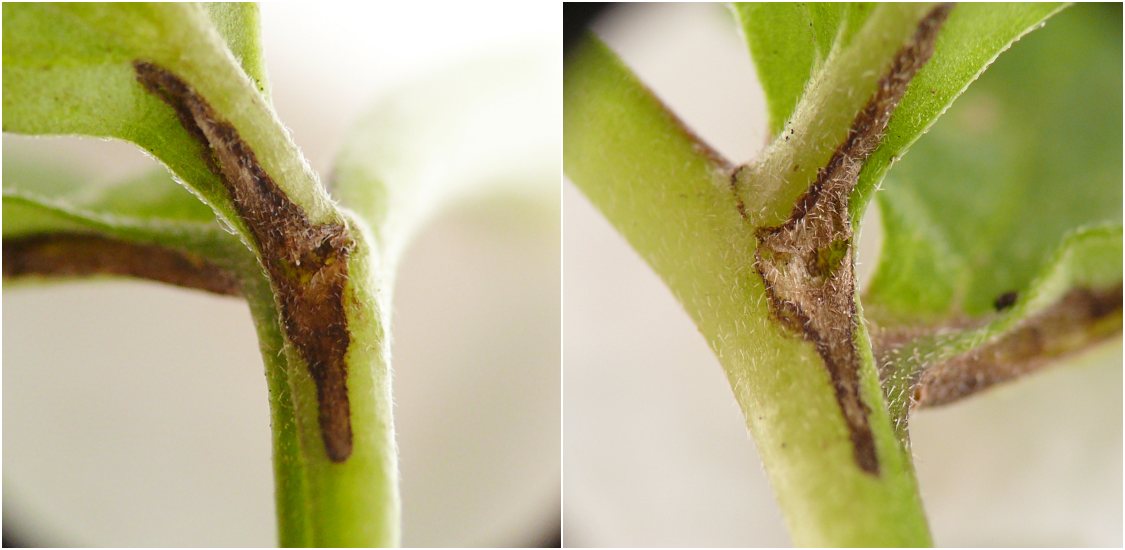
The epidermis on the upper surface of the petiole of this plant seems to be very loosely attached, and in several cases where an unknown force had caused a perforation in the epidermis, I found a linear cavity with darkened walls extending under the epidermis away from the perforation. In one case I observed a thrips exiting the petiole through the perforation, but was otherwise unable to associate any arthropod with the plant damage. As a result, the cavities in the images other than #30 and #33 below do not have a direct observational link to thrips, but I have included them nonetheless for the sake of comparison with the first two images.
Related observations
The following is a non-comprehensive assemblage of several additional publicly available reports of thrips living within enclosed areas in plants.
Hering (1951) described observations of thrips dwelling inside naturally-occurring cavities in plant tissue, and he pointed out that they do sometimes feed in these cavities, but he stopped short of calling this behavior "mining" unequivocally.
The possibility of members of [the order Thysanoptera] one day being discovered to be true miners is suggested by the fact that H. Buhr has found Thysanoptera in natural cavities of plants and their remnants of frass showed that some feeding had been taking place. Further investigations will have to determine whether at least in some cases true mining does occur in this Order. (p. 39)
[A]mong the species which are not so rigidly adapted to mining, there are some which feed in “natural” mines. They seek out natural cavities in the plant, in which they feed and sometimes remain for longer periods than would be expected from their normal life cycle. Such habits have been observed especially among Thysanoptera. (p. 264)
He also highlighted that thrips may be observed in mines of other insects. (The Dizygomyza example is credited to an unidentified "minologist.") Of particular interest here is the mention of epidermal holes established directly by the thrips:
[Feeding in secondary mines] can also be found among Thysanoptera, when the larvae enter mines which were constructed by other species and have already been abandoned by them. In this way they were found in old Dizygomyza mines on Gramineae. In these they constructed small, circular openings through which they ejected their frass... (pp. 264-265)
Eiseman (2014b) reported finding a species of Compsothrips inside small, bilobed cavities with a central perforation on agave leaves. He wrote, "Henry Hespenheide suggested that some of the round wounds might be feeding lesions of the conoderine weevil Peltophorus polymitus seminiveus (LeConte). He mentioned that Margarethe Brummermann has some nice photos of the weevil," which Eiseman found on Flickr, including an image of an adult standing next to some of the bilobed cavities on an agave plant (Brummermann 2011). Subsequent contributions to iNaturalist show adults of the same weevil species associated with fresh cavities and appearing to create them with their snouts, supporting this hypothesis (iNaturalist 2026b). However, the linear cavities with an entrance/exit hole on one end, which Eiseman observed on the agave leaves he examined and which were also later documented elsewhere by the iNaturalist photographers, evidently remain unexplained.
Eiseman (2014c) includes an image of a thrips visible through the outer wall of a silken cocoon spun by an unidentified insect on the surface of a leaf. Underneath the cocoon is a portion of a lepidopteran leaf mine, which has a small epidermal hole in it contiguous with the interior of the cocoon. Eiseman explained that "it seems like the thrips may have gotten trapped inside [the cocoon] after chewing its way out of the leaf mine."
Eiseman (2025) reviews instances in which thrips may appear in association with leaf mines, including the aforementioned observations referenced by Hering as well as a situation described by Arakaki and Okajima (1998) in which thrips preyed on agromyzid leafminer larvae without entering the agromyzid's mines. He also provides photographs on pages 48 and 49 depicting thrips from inside leaf mines, including a Eurythrips sp. from an agromyzid mine on honeysuckle and a "at least a dozen" thrips inside a chrysomelid mine on sea rocket. Also, Eiseman and Hannson (2026) document Ceranisus russelli (Eulophidae) reared from a thrips inhabiting a scathophagid leaf mine on Canada mayflower.
A compilation of images showing thrips found by several photographers inside galls; leaf mines or cavities; folded, curled, or rolled leaves; flowers; stems; and leaf sheaths is available on the BugGuide website (van der Linden 2026).
Discussion
I observed thrips inside cavities of several unrelated plants from 2022 through 2026. The internal blackening of the inhabited cavities may be a sign that the thrips are feeding on the inner walls of the cavities, but I was unable to confirm this. In cases where the inhabited cavity is positioned shallowly just underneath epidermis that is already loosely attached, it's tempting to ask whether the thrips could be enlarging the cavity somehow, even simply by wedging themselves further between the epidermis and the tissue underneath, thus leading to progressive separation of the epidermis, but I did not observe any such action directly. Hering (1951) asserts that "the construction of [thrips] mouth-parts would preclude the possibility of their producing a genuine mine," but he acknowledges that, when thrips inhabit abandoned mines of other insects, "[a]t least in some cases they consume parts of the leaf substance in the mine channel" (p. 290). Finally, the finding of an immobile prepupa or pupa inside a jewelweed petiole (see image 44) aligns with Hering's observation (as quoted in the "Related observations" section, above) that some thrips may stay inside these cavities for extended, developmentally relevant periods. Overall, these findings bolster observations made by previous workers (see, e.g., Hering 1951; Eiseman 2014b&c; Eiseman 2025; van der Linden 2026) that suggest a consistent association of thrips with cavities in plant tissue, a pattern worthy of further study in order to better elucidate the reasons for this behavior, its underlying mechanics, and its importance to the biology and ecology of the insects and plants involved.
References
- Arakaki, N. and S. Okajima. 1998. Notes on the biology and morphology of a predatory thrips, Franklinothrips vespiformis (Crawford) (Thysanoptera: Aeolothripidae): First record from Japan. Entomological Science 1(3): 359-363.
- Brummermann, M. 2011. Peltophorus polymitus seminiveus | Florida Canyon Santa Rita... | Flickr. Contributor post on Flickr. Retrieved July 24, 2026 from https://www.flickr.com/photos/margarethebrummermann/6042500455/in/photolist-acXoSk-9BQcdA/.
- Colorado State University. 2026. Thrips - Agricultural Biology. Integrated Pest Management (IPM) factsheet published by the College of Agricultural Sciences. Retrieved July 24, 2026 from https://agsci.colostate.edu/agbio/ipm-pests/thrips/.
- Eiseman, C.S. 2014b. Monthly mystery #14: holes and tunnels in agave leaves. BugTracks weblog. Retrieved May 28, 2026 from https://bugtracks.wordpress.com/2014/01/01/monthly-mystery-14-holes-and-tunnels-in-agave-leaves/.
- Eiseman, C.S. 2014c. Monthly mystery #21: double cocoons. BugTracks weblog. Retrieved May 29, 2026 from https://bugtracks.wordpress.com/2014/08/01/monthly-mystery-21-double-cocoons/.
- Eiseman, C.S. 2025. Leafminers of North America, 3rd edition. Self-published e-book. Available from the author at https://charleyeiseman.com/leafminers/.
- Eiseman, C.S. and C. Hansson. 2026. New rearing records for Eulophidae (Hymenoptera: Chalcidoidea) from the United States. Zootaxa 5826(1): 1-109.
- Hering, E.M. 1951. Biology of the leaf miners. Dr. W. Junk, publisher: The Hague. iv + 420 pp.
- iNaturalist. 2026b. Observations [of speckled agave weevil]. Retrieved July 24, 2026 from https://www.inaturalist.org/observations?taxon_id=304336.
- van der Linden, J. (ed.). 2026. Thrips inside plants. Contributor article at BugGuide.net. Retrieved July 24, 2026 from https://www.bugguide.net/node/view/2554901.